

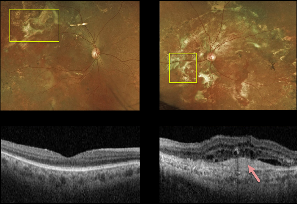
This 58YO male has stable vision of 20/20 OD and light perception OS. Extensive areas of subretinal fibrosis are scattered throughout each fundus, left greater than right.
A hyperreflective subfoveal scar on OCT in his left eye accounts for the poor vision. Fortunately, the right macula is relatively normal, and we are observing him twice yearly.
Learning Points:
Multifocal choroiditis (MFC) is the current preferred term for a group of historically heterogeneous disorders, including multifocal choroiditis, recurrent multifocal choroiditis, multifocal choroiditis and panuveitis, punctate inner choroidopathy, progressive subretinal fibrosis, and pseudo-histoplasmosis (Essex et al, Retina 2013;33:1-4 and Spaide et al, Retina 2013;33:1315-1324).
MFC occurs in otherwise healthy adults, often myopic and female. Findings include variably pigmented multifocal scars throughout the fundus, panuveitis, subretinal fibrosis, and macular neovascularization (MNV). Unlike ocular histoplasmosis, new scars and uveitis can be seen.
MNV can be successfully managed with anti-VEGF therapy.



