

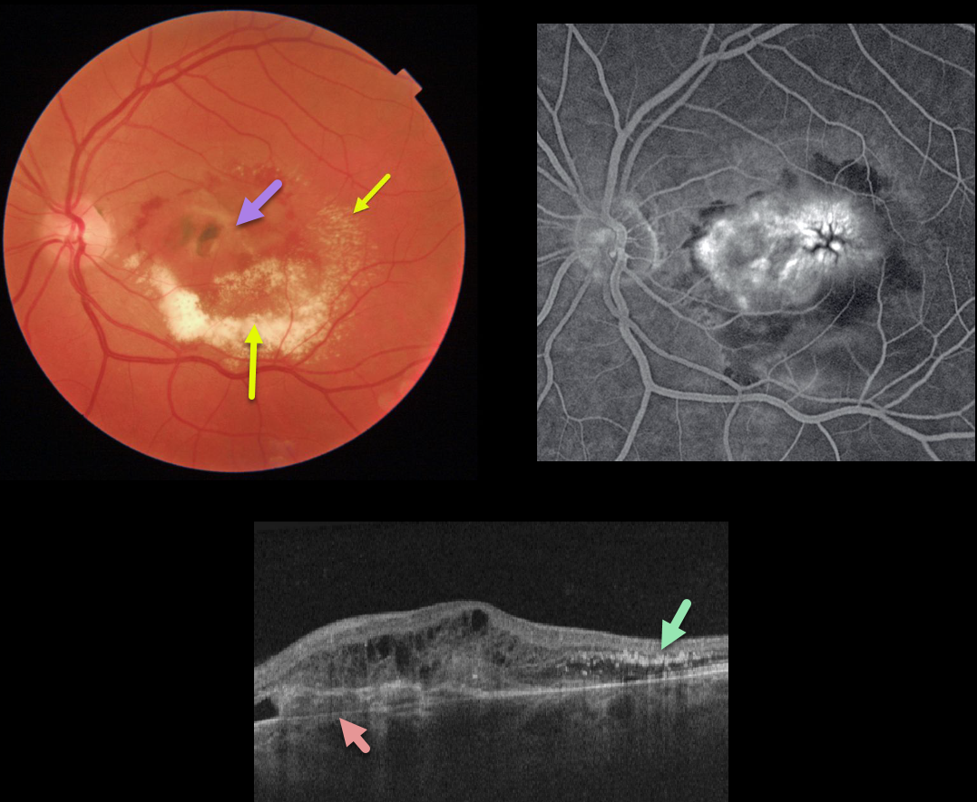
This patient was being followed for an inactive nasal macular chorioretinal scar with 20/25 vision. After being lost to follow-up for many months, he presented with counting-finger vision from an active type 1 macular neovascularization.
The macular OCT shows a complex lesion with the MNV located primarily beneath the RPE. The lipid precipitated out in the subretinal space and follows Henle’s layer temporally.
Learning Points:
The amount of lipid layered in the subretinal space is unusual in MNV. Extensive submacular lipid like this is more common with Coats disease or retinal capillary hemangiomas.



