

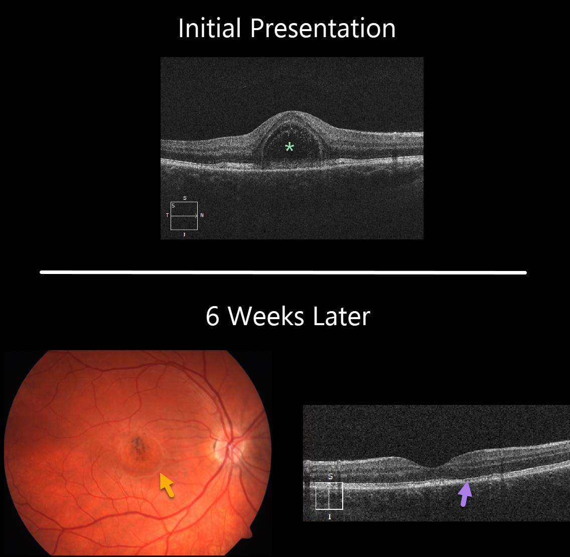
This 22YO female presented with 1 week of unilateral vision loss and 20/200 vision in her right eye.
The macular OCT line scan demonstrates a bacillary layer (cone and rod inner and outer segments) detachment. This appears as a unique dome shaped collection of intraretinal fluid from photoreceptor myoid splitting.
Six weeks later vision improved to 20/40. The macular fluid resolved, with the development of a dry area of macular hyperpigmentation surrounded by a rim of pigment loss. OCT shows some subtle outer retinal foveal thinning with ellipsoid zone irregularities.
Learning Points:
Acute idiopathic maculopathy (AIM) is a usually unilateral idiopathic condition usually found in young people, often following a viral illness. It is usually self-limited, resolving within several weeks with significant visual improvement. The appearance of a bulls-eye lesion, as occurred in our patient, is classic for this disease.
Bacillary detachments can be observed in multiple conditions including Vogt-Koyanagi-Harada (VKH) disease, AIM, blunt trauma, or APMPPE.
See Fernandez-Avellaneda et al Retinal Cases & Brief Reports 2019 (published ahead of print) for a discussion of bacillary layer detachment in AIM.



