

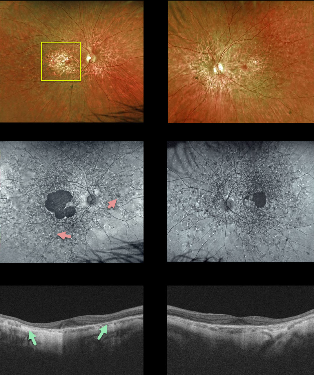
Our patient shows fairly classic findings of fundus flavimaculatus. Central vision remains good at 20/40 OD and 20/30 OS, with preserved foveas.
Findings include bilateral macular atrophy and scattered yellow, subretinal, multifocal, and autofluorescent flecks extending to the mid-peripheral retina. OCT shows RPE and outer retinal atrophy.
Learning Points:
Fundus flavimaculatus is a genetic disorder, usually transmitted as an autosomal recessive trait, that is caused by mutations involving the ABCA4 gene.
ABCA4 encodes one of a family of ATP-binding cassette (ABC) transmembrane proteins that mediate the active transport of various substrates across cellular membranes. The ABCA4 protein is present in the photoreceptor outer segment disc membranes, where it participates in the recycling of 11-cis-retinal.
Stargardt’s disease is felt to be fundus flavimaculatus without peripheral findings. Vision in fundus flavimaculatus is often better than Stargardt’s due to later disease onset and less macular involvement.
Both likely represent an RPE lipofuscin storage disease. The RPE cells become engorged with lipofuscin, causing a dark choroid on fluorescein angiography.



