

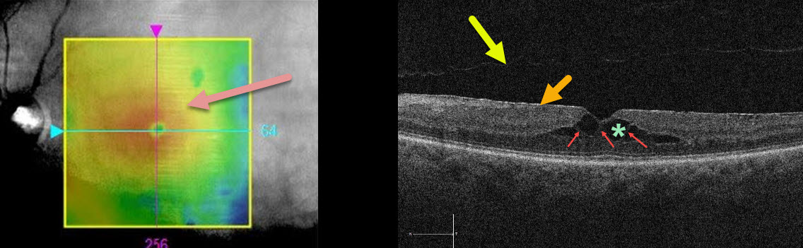
This patient was sent in for treatment for possible diabetic macular edema. The retinal thickness map shows central macular thickening.
The OCT B-scan shows an adherent, hyperreflective epiretinal membrane (ERM), a faint posterior vitreous detachment overlying the macula, and central cystic-like inner retinal spaces.
Muller cells, which extend vertically from the internal limiting membrane to the outer retina (external limiting membrane), essentially hold the 3 layers of retinal cells together.
In the central retina, the photoreceptor axons and Muller cells (Henle’s layer, which is the central OPL) are splayed outwards. This creates an intrinsically vertically weakened central macula. The central thin hyperreflective columns are Muller cells that span the region of split OPL.
Unlike exudative intraretinal cysts caused by leaking retinal blood vessels, these hyporeflective spaces are caused by tractional splitting of the outer plexiform layer (OPL) from either aborted vitreomacular traction and/or the ERM.
Learning Points:
When interpreting OCT scans, it is important to consider all the information in the OCT report, ideally by scrolling through the entire macular cube data in the review software.
The macula here is not truly edematous. It is thickened due to a tractional splitting of the retina. For a great discussion of tractional vs exudative cystoid spaces, see Govetto et al AJO 2020;212;43-56.



