

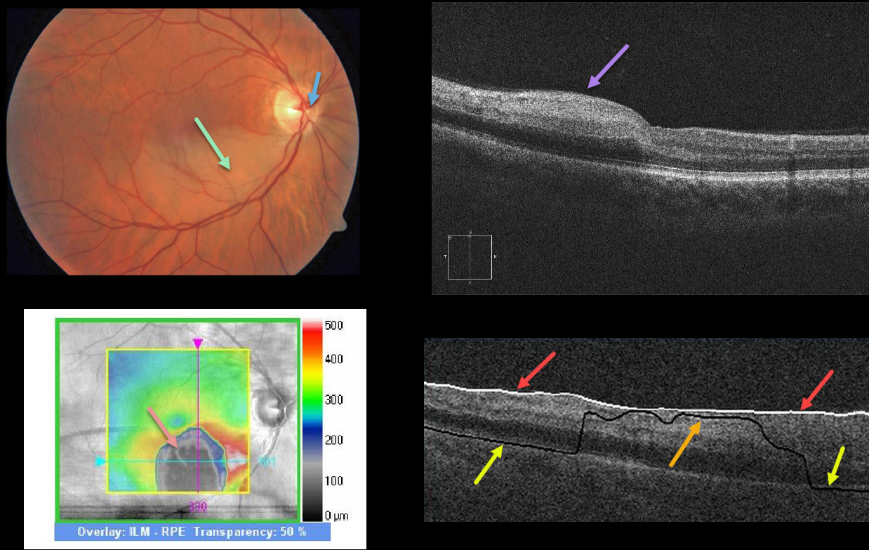
This patient has a subtle white, opaque retina in the inferior macula due to an acute branch retinal artery occlusion (BRAO). The causative embolus is the tiny white lesion just nasal to the nerve’s center.
OCT shows hyperreflectivity from the acutely ischemic inner retinal layers. The black blob on the retinal thickness map reflects an incorrectly identified RPE detachment (PED) caused by the algorithm misidentifying the RPE due to the marked hyperreflectivity of the inner retinal layers.
This phenomenon is demonstrated by the B-scan, which shows that the ILM is correctly identified by the white line. The black line is supposed to identify the RPE, which is correctly identified nasally and temporally but misidentified centrally due to said hyperreflectivity.
Learning Points:
An acute BRAO can be subtle clinically but more dramatic on OCT. Acute inner retinal OCT hyperreflectivity can lead to misidentification of retinal layers, resulting in artifacts.



