

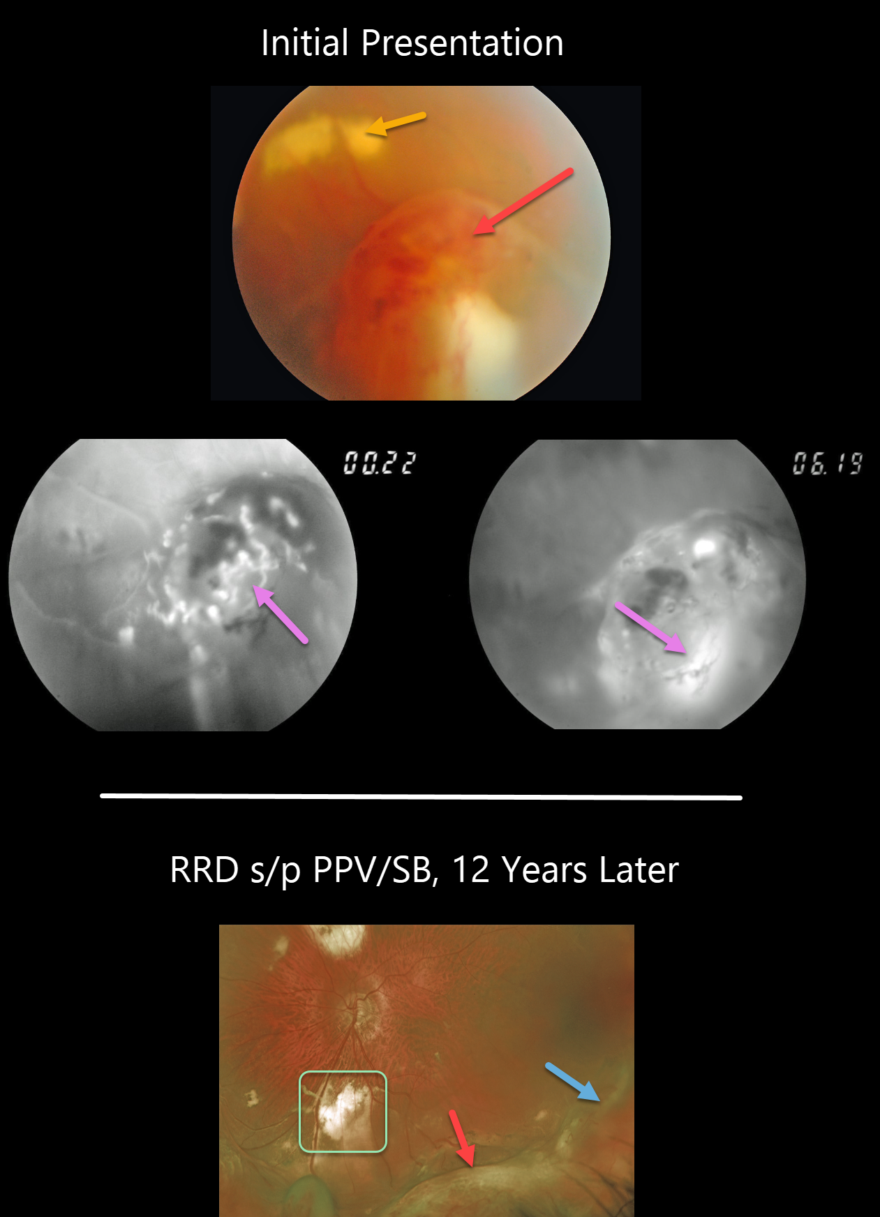
This 46YO presented with an asymptomatic vasoproliferative tumor (VPT). The VPT initially appeared as a tongue-like, reddish, elevated, peripheral mass with more posterior subretinal lipid. Fluorescein angiography (FA) revealed the vascular nature of the lesion.
He then developed a rhegmatogenous retinal detachment that was repaired with a combined vitrectomy and scleral buckle. The indentation from the buckle is visible more temporally and distinct from the complex enlarged VPT more inferiorly. The subretinal lipid resolved with more extensive subretinal scarring.
Learning Points:
Vasoproliferative tumors are benign globular, dome-shaped lesions arising in the peripheral retina. They can be yellow or red and cause subretinal exudation and detachment, anterior or posterior segment neovascularization, and vitreous hemorrhage.
They can be primary idiopathic lesions or a vascular response to various entities, including intermediate uveitis, retinitis pigmentosa, Coats disease, and prior retinal detachment.
For lesions causing symptomatic exudation or neovascularization, treatment with thermal laser, cryotherapy, and/or anti-VEGF injections is indicated.



