

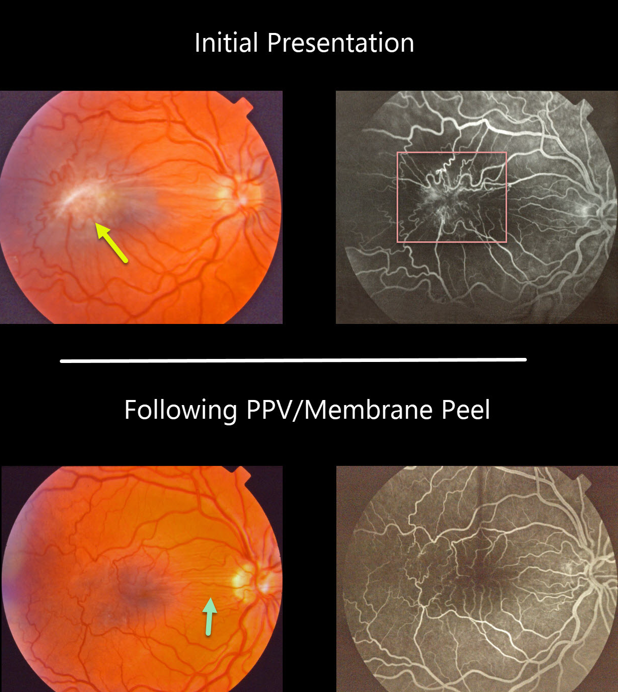
This patient had an isolated severe macular pucker. The fluorescein angiogram shows a vascular hamartomatous component due to severe retinal vascular distortion and leakage. This vascular component, along with the pigmented appearance of the pucker, helps give the false appearance that this was a combined hamartoma of the RPE.
The macular pucker was treated with vitrectomy and membrane peeling. Postoperatively, the retina appears nearly normal, with some residual peripapillary retinal folds and no vascular leakage.
Learning Points:
Hamartomas and severe macular puckers can look very similar, even to some of the most seasoned retina specialists. OCT may be helpful in differentiating these entities (see Ophthalmic Surg Lasers Imaging Retina 2017;48;122-125).



