

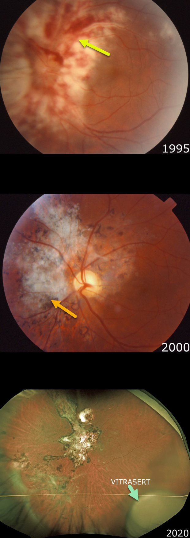
Our patient presented in 1995 with classic active CMV retinitis involving the peripapillary and macular regions of his left eye. We immediately referred him to the University of Kentucky for implantation of a then-investigational sustained-release ganciclovir implant, Vitrasert.
Note the hemorrhagic, whitish retinal necrosis, which often follows a vascular distribution. The retinitis spreads outwards, leaving behind a thinned necrotic retina and RPE.
Following Vitrasert implantation, the retinitis resolved with secondary scarring. He was then started on highly active antiretroviral therapy (HAART).
In 2007, a rhegmatogenous retinal detachment, a common late sequela of CMV infection, was successfully repaired with vitrectomy (not pictured).
As of 2020, he continues to do well with an undetectable viral load and a normal CD4 count. Vision is 20/20 in his normal right eye and 20/400 OS. The ganciclovir Vitrasert implant remains visible in the inferotemporal periphery.
Learning Points:
Cytomegalovirus (CMV) retinitis develops as a reactivation of latent CMV in immunosuppressed individuals. Before effective antiretroviral treatment emerged in the mid to late 1990’s, CMV retinitis developed in up to 40% of HIV/AIDS patients, often within the last 6 months of life.
Treatment includes a combination of intravenous and intravitreal medications, including ganciclovir, foscarnet, and cidofovir.
Vitrasert was approved by the FDA in 1996 and discontinued in 2013 due to patent expiration. The demise of Vitrasert is directly linked to HAART.
As occurred in our patient, pharmacologic restoration of immune function allows the patient’s own immune system to control CMV.



