

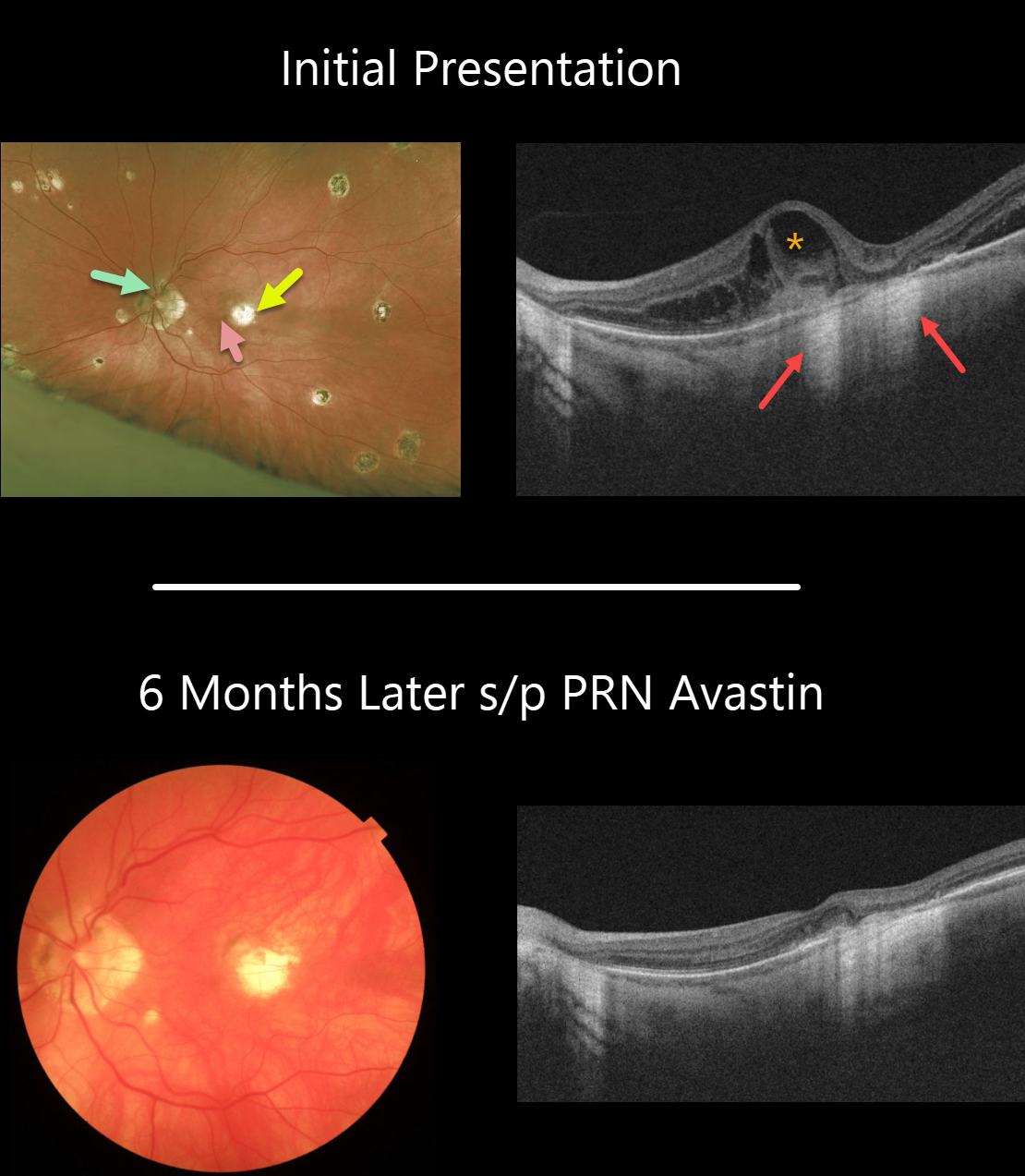
Our patient has 20/20 vision OD and 20/400 OS. He has classic peripapillary atrophy, macular and peripheral scarring.
He came in with symptomatic vision loss in his left eye with a subtle nasal foveal subretinal hemorrhage from an acute macular neovascularization (MNV) adjacent to a pre-existing chorioretinal scar.
The OCT line scan surprisingly shows significant intraretinal fluid overlying the hemorrhage and temporal to the clinical scar. The loss of the RPE at the chorioretinal scar allows OCT to show increased hyperreflectance of the underlying choroid and sclera. This is the opposite of shadowing.
Six months later, vision in the left eye is 20/200, and the blood and fluid remain completely resolved with PRN Avastin.
Learning Points:
Ocular histoplasmosis is caused by the Histoplasmosis capsulatum fungus, which is commonly found in soil contaminated by bird or bat droppings. It is found worldwide, but in the United States, it is most common in the Ohio-Mississippi River valleys.
The vast majority of people in endemic areas inhale the organism’s spores early in life, and only a few percent will develop asymptomatic chorioretinal scars. A small percentage of these will develop MNV later in life.
Originally described by Woods and Wahlen in 1959, the findings include the classic triad of peripapillary atrophy, peripheral multifocal punched-out scars, and a macular scar or neovascularization.
We like to add a fourth criterion: a quiet vitreous, since numerous inflammatory disorders can simulate ocular histoplasmosis.



