

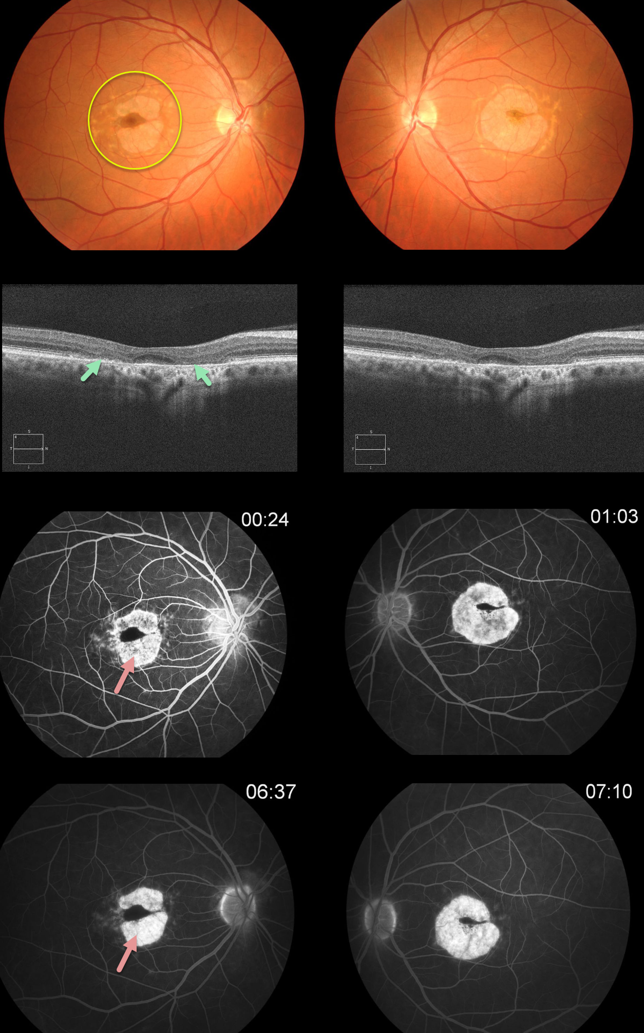
This patient has classic findings for Plaquenil (hydroxychloroquine) toxicity, including a bull’s-eye pattern of pericentral RPE and outer retinal atrophy. This is seen clinically as loss of the orange RPE pigment, on OCT as loss of the photoreceptor elements and RPE, and on fluorescein angiography as hyperfluorescent window defects.
In the pre-OCT era, patients were followed until clinical or angiographic pigmentary changes developed, at which point the medication was discontinued. Unfortunately, this patient was not properly screened and developed irreversible changes.
Learning Points:
Current screening guidelines should make Plaquenil toxicity a disease of the past
Optical coherence tomography (OCT) is performed annually to assess outer retinal findings, including ellipsoid zone loss.
Humphrey 10-2 visual field testing (24-2 for Asians, since their macular involvement is usually more peripheral) is also needed annually, as about 10% of patients will have field loss despite normal examinations and OCT testing. Plaquenil dosing should also be based on real, not ideal, weight to better predict the optimal dose.
See Marmor et al, Ophthalmology 2016;123:1386-1394 for the full screening guidelines.



