

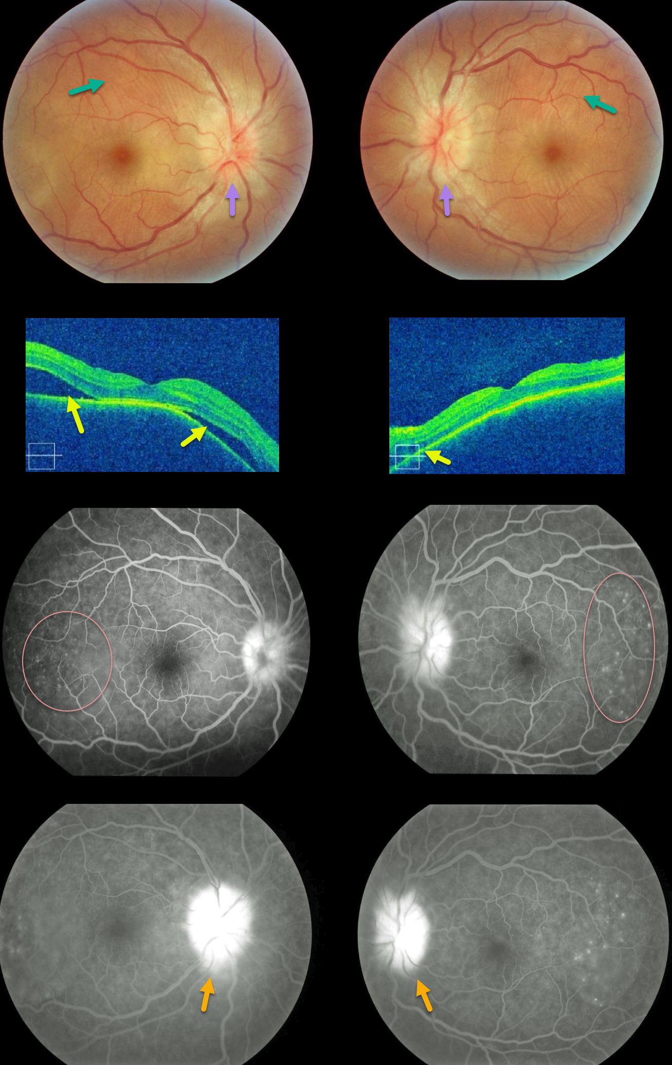
Our patient shows classic findings for acute Harada’s. The fundus photos and OCT images show bilateral multifocal subretinal exudative detachments with irregular, scattered overlying chorioretinal folds and bilateral optic disc swelling.
Fluorescein angiography (FA) shows multifocal subretinal leaks with pooling of fluid below the retina and within outer retinal cystoid spaces, along with leaking “hot” nerves.
These findings dramatically resolved after just 1 week of oral prednisone (not pictured).
Learning Points:
Bilateral, multifocal yellow-white, serous retinal detachments are the hallmark sign of acute Vogt-Koyanagi-Harada (VKH) disease.
The choroid is markedly thickened with overlying chorioretinal folds. The multiple sets and various orientations of these folds are quite different from those seen with idiopathic chorioretinal folds.
The optic nerves are usually inflamed and swollen. Patients often have a panuveitis with anterior chamber and vitreous cells.
Those with just ocular findings have Harada disease, whereas patients with additional systemic findings (including vitiligo, poliosis, headache, vertigo, and hearing loss) have Vogt-Koyanagi-Harada disease.



