

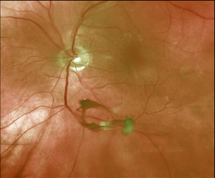
This 67YO male presented with a symptomatic posterior vitreous detachment (PVD). A prominent Weiss ring is beautifully captured in this Optos image.
His peripheral retina was normal, and he was asked to return in a few months unless he developed new flashes, floaters, or vision loss.
Learning Points:
The clinical diagnosis for a complete PVD can be established with biomicroscopy when a Weiss ring, representing avulsed peripapillary glial tissue, is visualized. OCT can be helpful in supplementing the clinical exam (see Wagley et al, Retina 2021;41:2296-2300).
An acute PVD is the moment of truth for when a retinal break or detachment develops. The historical risk for a break is at least 10-15%, although a recent large retrospective study from Kaiser Permanente found this risk to be significantly lower at 5% for a retinal break and 4% for retinal detachment (Ophthalmology 2022;129;67-72).
The presence of vitreous pigment or blood and lattice, and a history of a retinal break or detachment in the fellow eye, increases this risk.



