

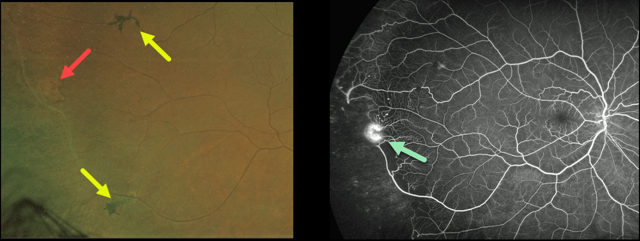
This 56YO African American male presented with floaters in his right eye and a history of sickle cell anemia. Vision was 20/30 OU.
A mild vitreous hemorrhage was noted inferiorly, along with an area of suspected peripheral retinal neovascularization. There were several areas of midperipheral black sunburst lesions. Fluorescein angiography confirmed a patch of retinal neovascularization at the junction of perfused and non-perfused retina. Due to the symptomatic vitreous hemorrhage, scatter laser to the peripheral ischemic retina was recommended.
Learning Points:
Patients with sickle cell disease develop symptoms throughout their bodies as stiff, sickled red blood cells block capillary beds. In the eye, this can cause proliferative sickle cell retinopathy (PSR) with sea-fan-shaped areas of peripheral retinal neovascularization developing at the junction of perfused and ischemic retina.
The black sunburst lesions are thought to occur from intraretinal RPE migration in response to photoreceptor damage from intraretinal and subretinal hemorrhage (see Ausdourian et al, BJO 1975;59:710-716). Complications include vitreous hemorrhage and traction retinal detachment. Although patients with SS disease tend to have more systemic complications, SC disease is usually associated with more severe ocular findings.
Treatment with peripheral scatter laser photocoagulation is somewhat controversial because, unlike in more typical proliferative retinopathies (e.g., those from diabetes and branch retinal vein occlusions), these new vessels tend to auto-infarct.
For a great review article on sickle cell disease and the eye, see Elagouz et al, Surv Ophthalmology 2010;55:359-377.



