

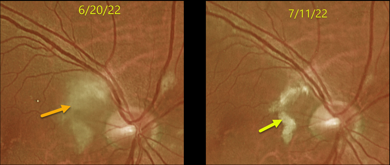
This 65YO male developed inferior field loss in his right eye immediately following cardiac valve replacement on 6/10/22. Vision was 20/30 when he presented to our office 10 days later.
Optos imaging shows patches of inner retinal opacification in the nasal macula and around the nerve, consistent with Purtscher ’s-like retinopathy. Several weeks later, the opacification was resolving with improving symptoms.
Learning Points:
Purtscher’s retinopathy was first described by Otmar Purtscher in 1910. Although originally described in a man who fell from a tree with cranial trauma, these white patches of retinal ischemia and hemorrhages are found in numerous other etiologies, including pancreatitis, chest trauma, and collagen vascular diseases.
The findings are called Purtscher’s retinopathy when due to trauma, and Purtscher-like retinopathy when seen from other causes. The underlying cause of Purtscher’s is unknown, but it is thought to represent leukoembolization by complement activation. The areas of ischemia include more superficial nerve fiber layer infarcts (cotton wool spots) and deeper ischemia involving the middle retinal layers.
This middle retinal ischemia, paracentral middle maculopathy (PAMM), is a recently-described phenotype found as an idiopathic entity or in other ocular disorders, including Purtscher’s, diabetic retinopathy, retinal artery and retinal vein occlusions, and acute macular neuroretinopathy (see Retina 2015;35:1921-1930).



