

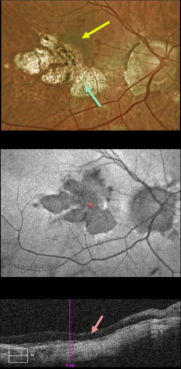
This 51YO female received prior intravitreal injections in her right eye about 5 years earlier, but was subsequently lost to follow-up until she was referred to our office. She felt that her vision had been stable and poor in each eye for years. Vision was counting fingers OD and 20/200 OS.
Optos color imaging of her right eye shows myopic peripapillary and macular scarring extending through the macular center. A pigmented macular neovascularization (MNV) with some dots of subretinal blood extends superonasally from the atrophy. Fundus autofluorescence (FAF) shows areas of hypo-FAF corresponding to the areas of clinical atrophy. Triton swept-source OCT shows central retinal atrophy.
Given the foveal atrophy, severe vision loss, and absence of acute symptoms, we recommended careful observation. We will consider anti-VEGF therapy if the MNV causes increasing exudation.



