

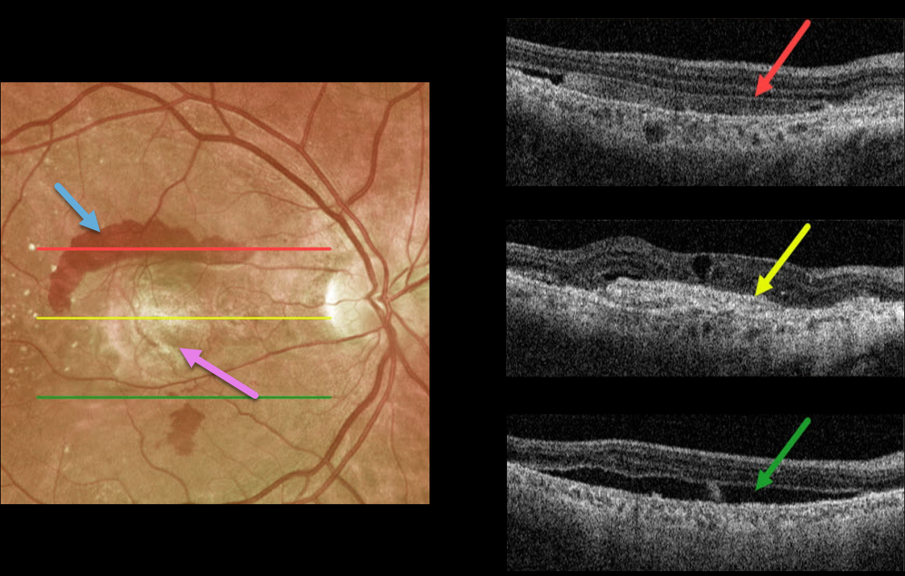
This 72YO male presented with longstanding decreased vision in his right eye. Vision was counting fingers due to a fibrosed scar from neovascular age-related macular degeneration (nAMD).
Optos imaging shows a central fibrosed macular scar with superior and inferior subretinal blood. Triton swept-source imaging shows multiple cool findings from this single neovascular lesion. Superiorly, the subretinal blood appears as hyperreflective material in the subretinal space. More centrally, the fibrotic scar/type 2 macular neovascularization is hyperreflective. There are also some overlying chronic cystic retinal changes. More inferiorly is shallow, subclinical subretinal fluid.
A therapeutic trial of monthly anti-VEGF injections was initiated, although we’re not sure this will have a visual effect given the subfoveal fibrosis.



