

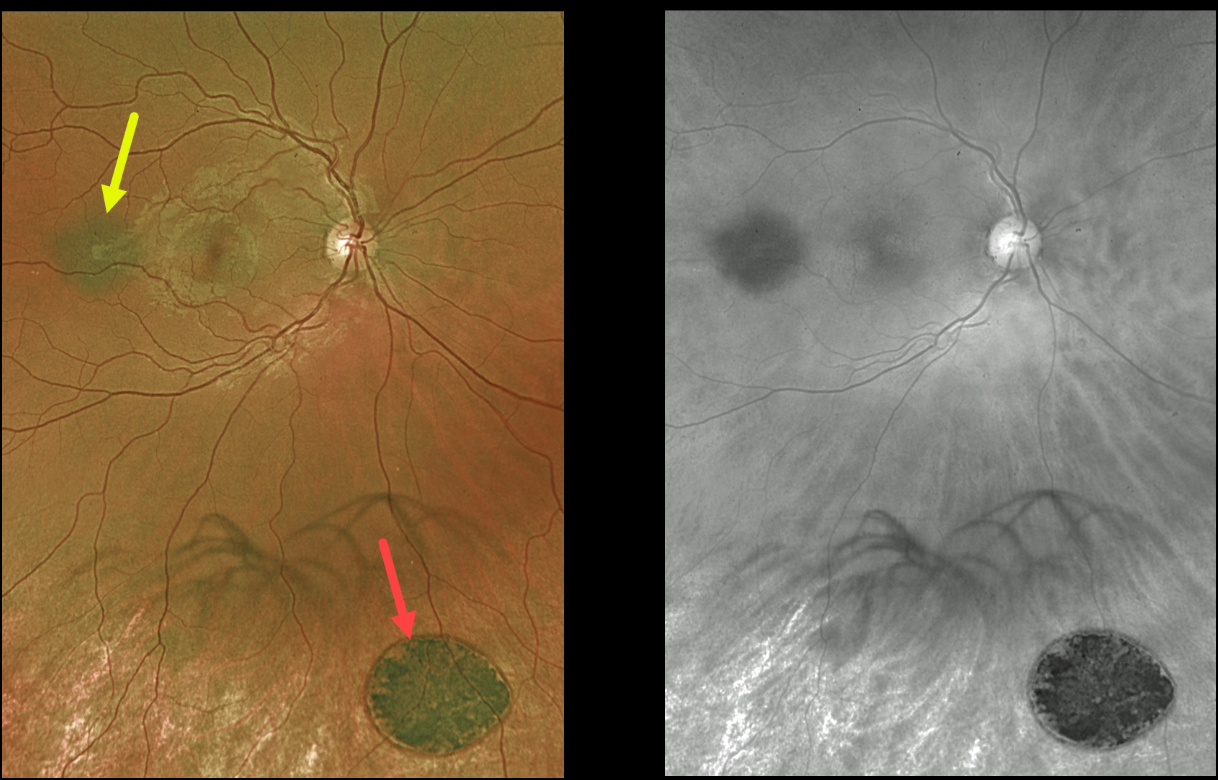
This 27YO male was referred for two distinct and asymptomatic pigmented fundus lesions.
Optos color imaging shows a small, flat choroidal nevus and a larger, flat patch of congenital hypertrophy of the RPE. The color image best shows the color difference between these lesions, with the CHRPE lesion appearing jet-black compared with the nevus.
The red channel best highlights the typical, somewhat indistinct margins of the nevus compared with the very sharp, round margins typical for the CHRPE. The patient is being followed yearly.
Learning Points:
Clinical characteristics of choroidal nevi at risk for progressing to a uveal malignant melanoma were initially described by Shields et al (Ophthalmology 1995;102:1351-1361).
The most recent version of this famous acronym (see Retina 2019;39:1840-1851) is To Find Small Ocular Melanoma Doing Imaging (TFSOM-DIM), which stands for:
To (Thickness >2mm)
Find (subretinal Fluid)
Small (Symptoms, vision <= 20/50)
Ocular (Orange pigment)
Melanoma (Melanoma hollow on ultrasonography)
DIM (DIaMeter > 5mm)
Although CHRPE lesions can enlarge with time (see Shields et al, Ophthalmology 2003;110:1968-1973), they carry virtually no malignant potential.
Histologic RPE hypertrophy presents in several ways. The typical isolated CHPRE lesions and bear tracks are not associated with intestinal polyps or cancer, and thus require no systemic evaluation. Only the multiple, small comet-shaped lesions are part of the familial polyposis spectrum.



