

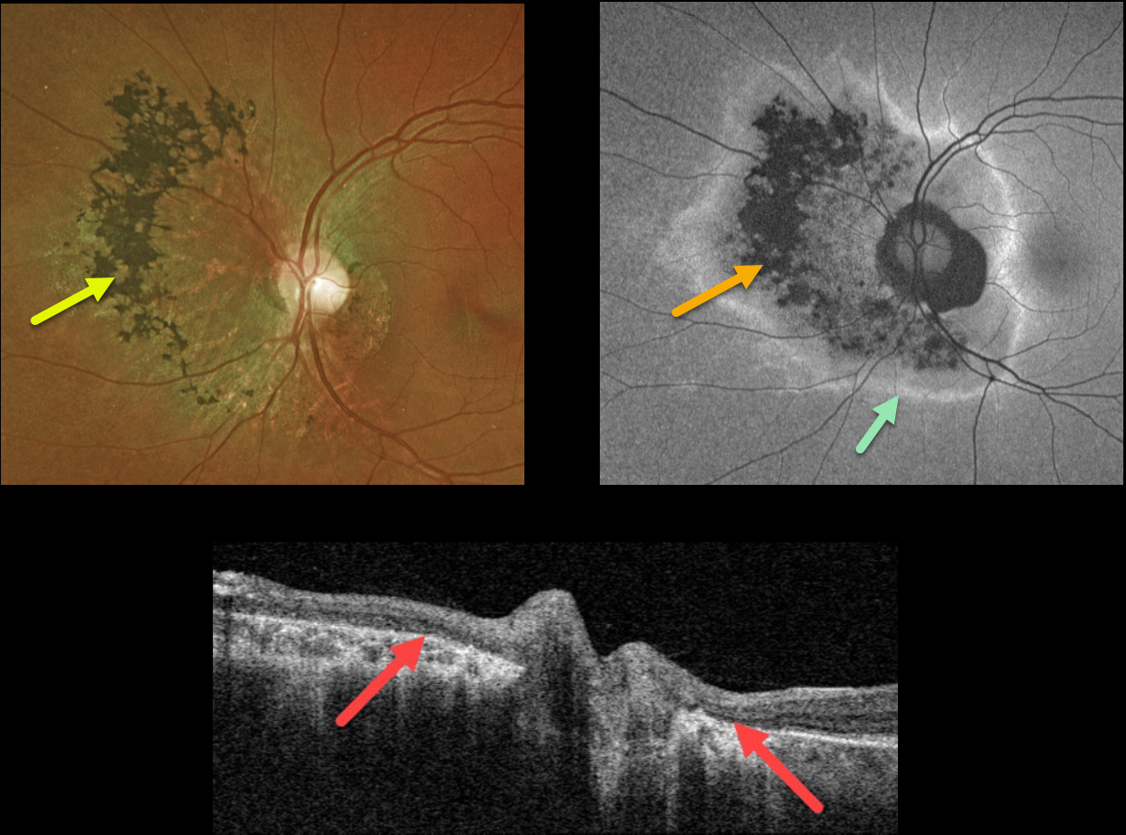
This 44YO female was referred for asymptomatic retinal changes in her right eye. Vision was 20/20 in her normal right eye and 20/30 OS.
Optos color imaging shows chorioretinal scarring surrounding the left optic nerve, extending more nasally with intraretinal pigment migration. Fundus autofluorescence (FAF) shows hypo-FAF corresponding to the clinical chorioretinal scarring, with a surrounding rim of hyper-FAF. Swept source OCT through the optic nerve shows loss of the outer retinal bands with RPE atrophy.
We made a presumed diagnosis of resolved acute zonal occult outer retinopathy (AZOOR)
Learning Points:
First described by Gass in 1992, AZOOR initially presents unilaterally or bilaterally in young women with photopsias, central visual changes, and minimal fundoscopic changes. Zones of visual field loss correspond to the locations of the photopsias. Electroretinography is often abnormal. Some may later develop areas of outer retinal, RPE, and choroidal atrophy in the initially affected retina.
The pathogenesis of the condition remains unclear, but it appears to initially involve inflammation of the photoreceptors. AZOOR is believed to be a part of the spectrum of other ‘photoreceptoritis’ conditions, including multiple evanescent white dot syndrome (MEWDS), acute macular neuroretinitis (AMN), and acute retinal pigment epitheliitis.



