

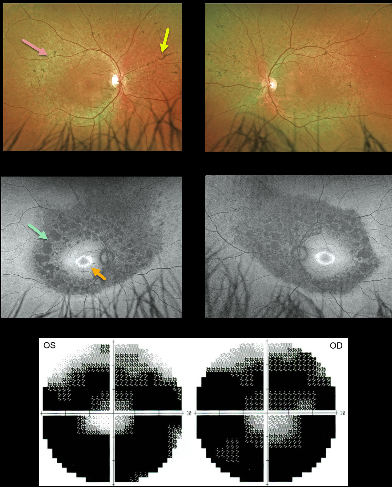
This 34YO male was referred for relatively asymptomatic retinal findings. He had complained of some blurred distance vision for several years. Vision was 20/25 OU.
Optos color imaging shows fairly normal maculas with midperipheral encircling pigment loss and intraretinal pigment migration. Fundus autofluorescence (FAF) shows a bull’s-eye-shaped area of macular hyper-FAF, and areas of midperipheral and nasal hypo-FAF corresponding to the pigmentary changes clinically. Visual field testing shows marked bilateral constriction. Invitae genetic testing was negative.
Learning Points:
Patients with retinitis pigmentosa (RP) classically have pale optic nerves, narrowed retinal blood vessels, variable outer retinal and RPE loss, and intraretinal pigment migration (bone spicules) due to loss of the inhibitory effect of the degenerated outer segments, allowing the RPE to migrate along the retinal capillaries.
A negative genetic test does not mean this patient doesn’t have RP; rather, it reminds us that such testing must be interpreted with caution and always with the clinical findings in mind.



