

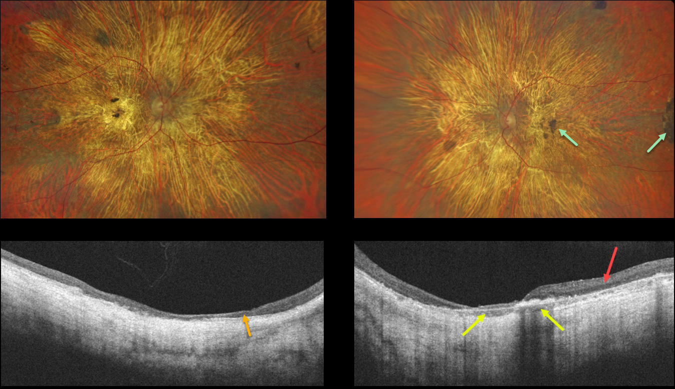
This 56YO male presented with a long history of vision loss and night blindness. Vision was counting fingers bilaterally.
Optos imaging shows severe, bilateral diffuse chorioretinal atrophy with some scattered large clumps of pigmentation. OCT scanning of the right eye shows diffuse thinned, disorganized retina and no choroidal structures. OCT scan of the left eye shows relatively preserved temporal retina and some preserved choroid.
Learning Points:
Choroideremia is an X-linked recessive chorioretinal dystrophy caused by a mutation in the CHM gene located in the long arm of the X chromosome. Sons of female carriers have a 50% chance of developing choroideremia, and daughters have a 50% chance of becoming carriers. Some female carriers can still develop choroideremia due to irregular inactivation of the X chromosome (see Jauregui et al, AJO 2019;207:77-86).
Night blindness usually begins between the ages of 10 and 30, followed by peripheral visual field loss. Zones of patchy RPE and chorioretinal atrophy initially appear in the mid-periphery, gradually spreading anteriorly and posteriorly. Eventually, the patient is left with only a small island of vision, narrowed retinal vessels, and optic atrophy.
For a great choroideremia review article, see Pennesi et al., Retina 2019;39:2019-2069.



