

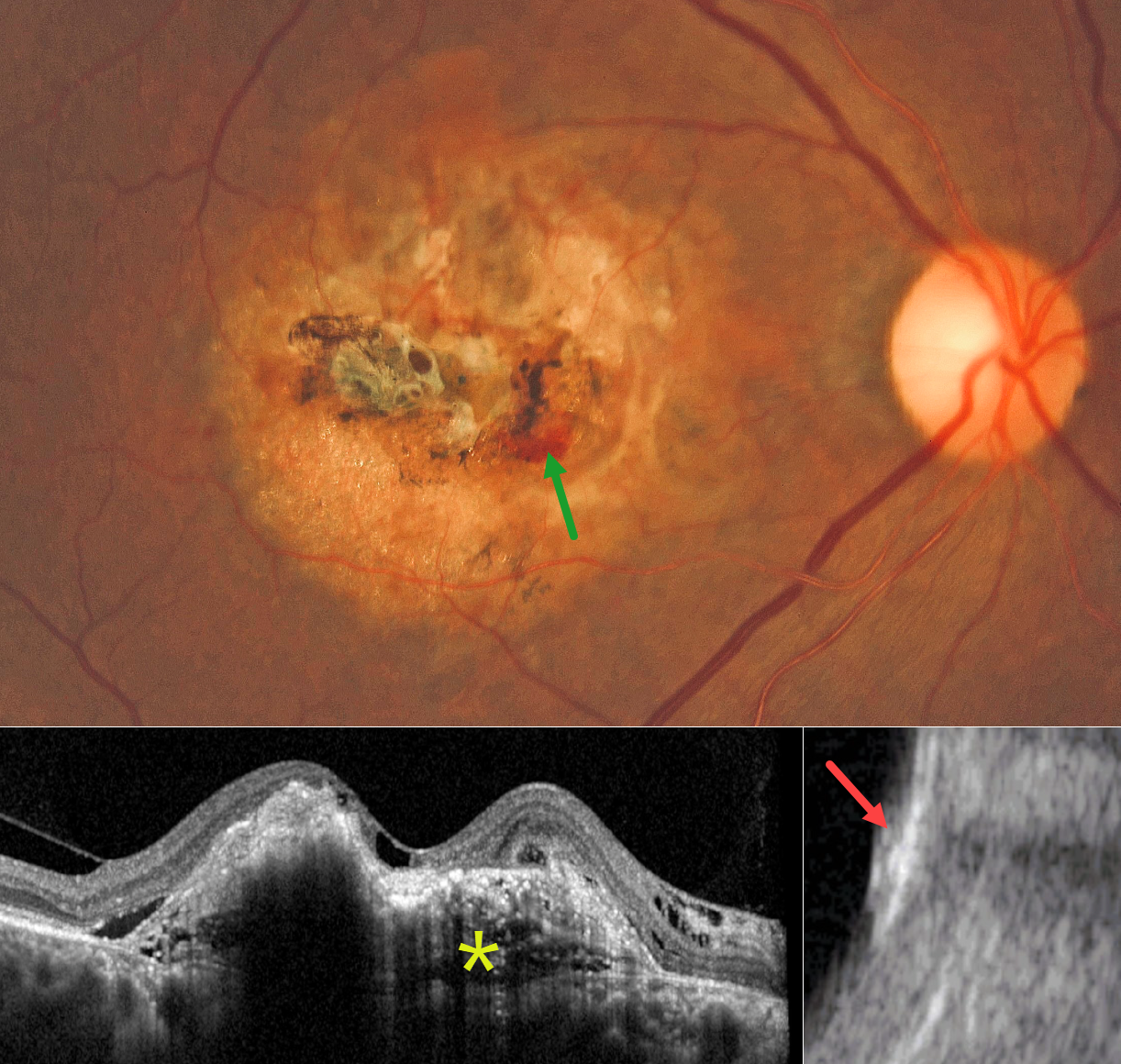
This 73YO male presented with 8 months of gradual vision loss in his right eye. Vision was 20/200 OD and 20/25 in his normal OS.
Color photography shows a yellowish, refractile amelanotic occupying nearly the entire macula. Some areas of increased pigment are noted centrally, along with some blood inferonasally. On OCT, the lesion has an internal lamellar, sponge-like architecture, characterized by horizontal hyporeflective lines and some intraretinal and subretinal fluid. The lesion is markedly hyperreflective on B-scan ultrasonography with posterior acoustic shadowing.
Learning Points:
Choroidal osteoma is a rare, benign ossifying choroidal tumor that typically affects young women, often in the peripapillary or macular region. Typical features include a clinical yellowish-orange lesion, a hyperreflective choroidal mass with a lamellar or trabecular internal architecture on OCT, and, most importantly, marked posterior acoustic shadowing on B-scan ultrasonography due to calcification. These imaging characteristics are distinct from those of other choroidal lesions, such as melanoma or hemangioma, which lack dense calcification and exhibit different reflectivity patterns.
Although benign, choroidal osteoma may cause vision loss due to macular neovascularization (MNV), subretinal fluid, and progressive decalcification, with atrophy of the retinal pigment epithelium and outer retinal layers. MNV can be successfully treated with anti-VEGF therapy (see Seong et al, Graefe’s 2022;250:1713-1721). Intravitreal Eylea was started for our patient.



