

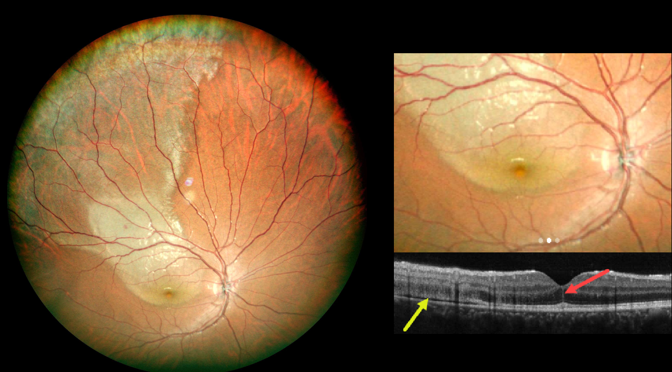
This 15YO boy presented 1 day following blunt injury to his right eye from a soccer ball. Despite complaints of a central scotoma, vision was 2025.
Color photography shows a broad area of commotio retinae extending from the central macula into the superotemporal periphery. OCT scanning shows disruption of the outer retinal bands and shallow subretinal fluid temporally. A vertical hyperreflective line is present in the central outer nuclear layer.
Learning Points:
The foveal crack sign (FCS) is a central foveal hyperreflective vertical line, first described by Ishibashi et al. as an OCT biomarker for future macular hole development following vitrectomy for rhegmatogenous retinal detachment (AJO 2020;218:192-198). It has since been described in numerous other conditions, including macular pucker, diabetic macular edema, idiopathic macular telangiectasia type 2, vitreomacular traction, NAION with CRAO, AMD, non-infectious uveitis, Coats disease, and commotio retina (Kayabasi et al, Med Hypothesis Discov Innov Ophthalmol 2024;13:129-138).
Commotio retinae is caused by direct, blunt eye trauma. The outer retinal whitening is due to shearing of the photoreceptor outer segments. The term “Berlin’s edema” is a misnomer and should not be used, as the retina is not edematous. The acute white retinal changes usually resolve within several weeks. With resolution, the fundus appearance can normalize. With more severe injury, outer retinal loss can allow RPE cells to migrate into the retina, giving a pseudo-retinitis pigmentosa appearance. Patients can also develop retinal breaks and detachment.
We will follow our patient closely and hope that the FCS is not an omen for a future macular hole.



