

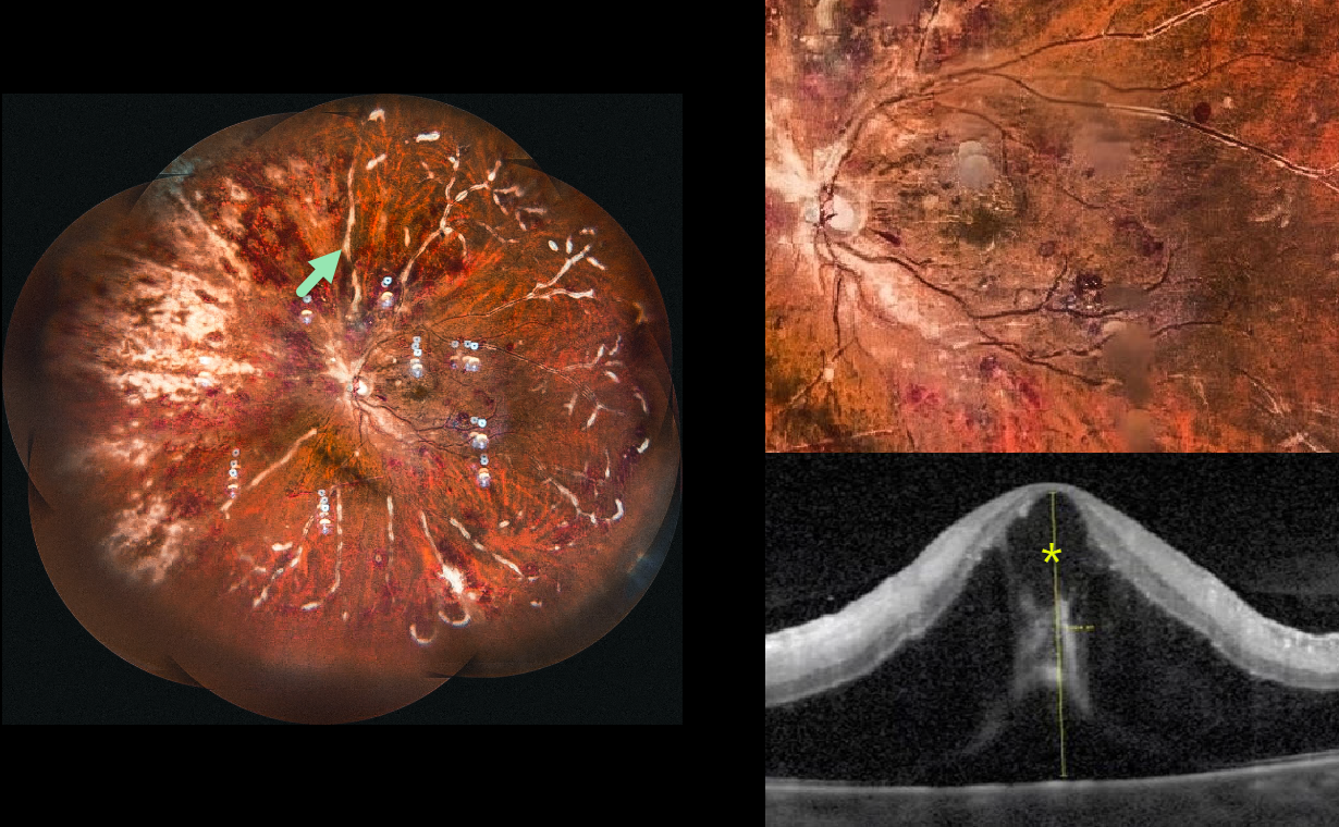
This 45YO female with a known history of poorly controlled systemic lupus erythematosus (SLE) presented to us for a third opinion with 10 days of bilateral vision loss. Vision was 20/100 OD and 20/200 OS.
Ultrawidefield imaging of her left eye shows extensive retinal hemorrhages with a frosted branch appearance to the retinal veins. Marked cystoid edema and subfoveal fluid are noted on OCT. Identical findings were seen in her right eye (not shown). She was immediately lost to follow-up.
Learning Points:
Frosted branch angiitis is a retinal vasculitis that gets its name from the involved vessels resembling branches of a tree that are “frosted” with snow (Kleiner et al, AJO 1988;106:27-34). Although these findings may be isolated, they are often considered a phenotype found in many inflammatory conditions, most commonly CMV retinitis. Inflammation is usually highly responsive to steroids, and the visual prognosis is generally good, although some may develop secondary neovascularization in the anterior and posterior segments due to widespread ischemia.
Although frosted branch angiitis has not been specifically linked to SLE, retinal vasculitis (arteriolar vasculitis with cotton wool spots, retinal hemorrhages, and vascular occlusions) is a known lupus finding (Gao et al, Lupus 2017;26:1182-1189).



