

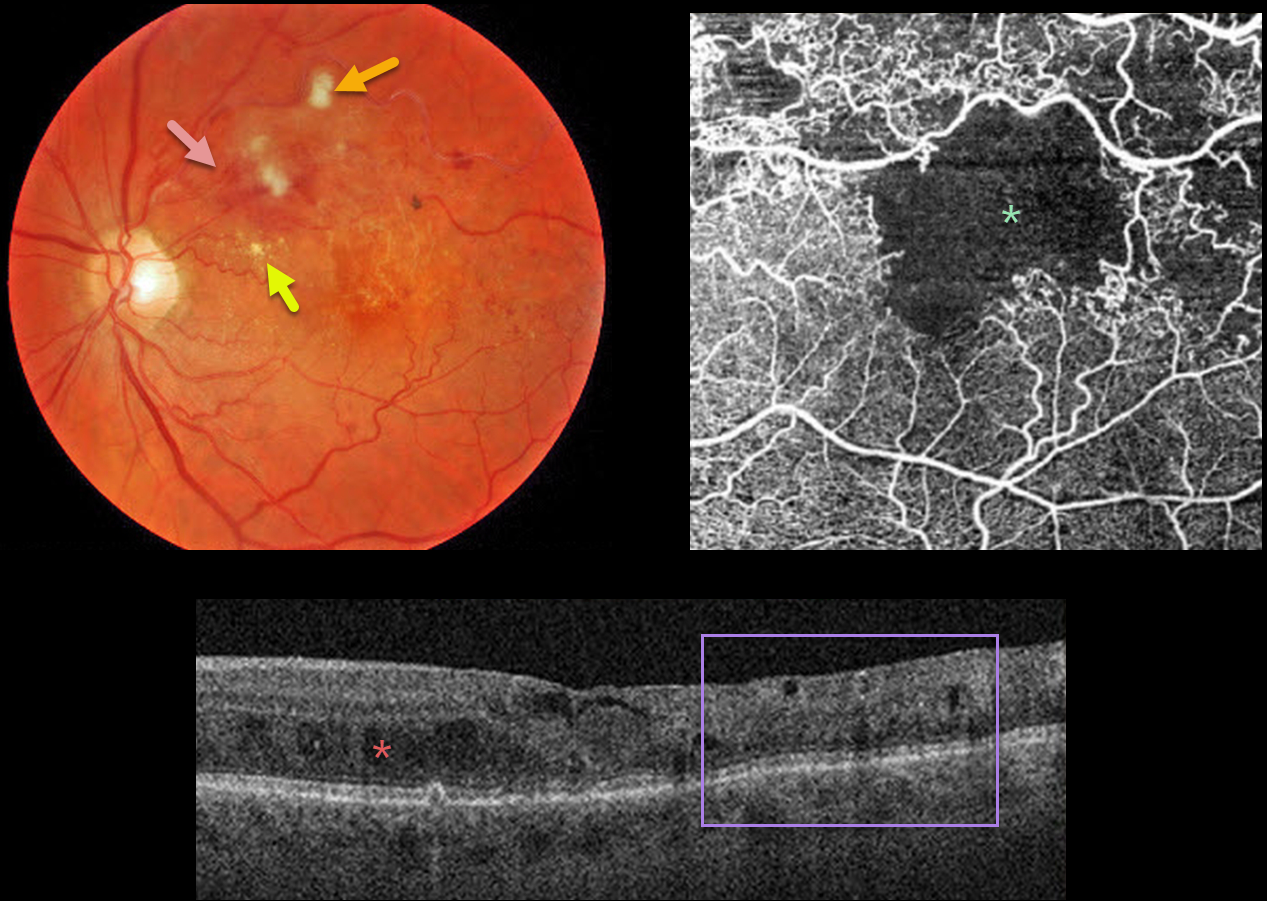
This 79YO male presented with a major superotemporal branch retinal vein occlusion (BRVO) and counting fingers vision.
Fundus photography shows superior macular hemorrhages with inner retinal ischemia (cotton-wool spots) and some nasal lipid. The vessels, especially around the superotemporal fovea, are nonperfused.
The clinically suspected foveal ischemia is confirmed on OCT angiography. Spectral-domain OCT shows disorganization of the inner retinal layers (DRIL), particularly within the clinically ischemic temporal macula, along with scattered inner and outer retinal cysts.
Learning Points:
DRIL is seen in various retinal vascular disorders, including diabetic macular edema and retinal vein occlusions. It is generally associated with worse vision and increased anti-VEGF treatment burden (see Babiuch et al, JAMA Ophthalmol 2019;137:38-46).
Since the patient has no central foveal thickening or retinal neovascularization, we are observing for now. Anti-VEGF therapy will not restore the foveal circulation.



