

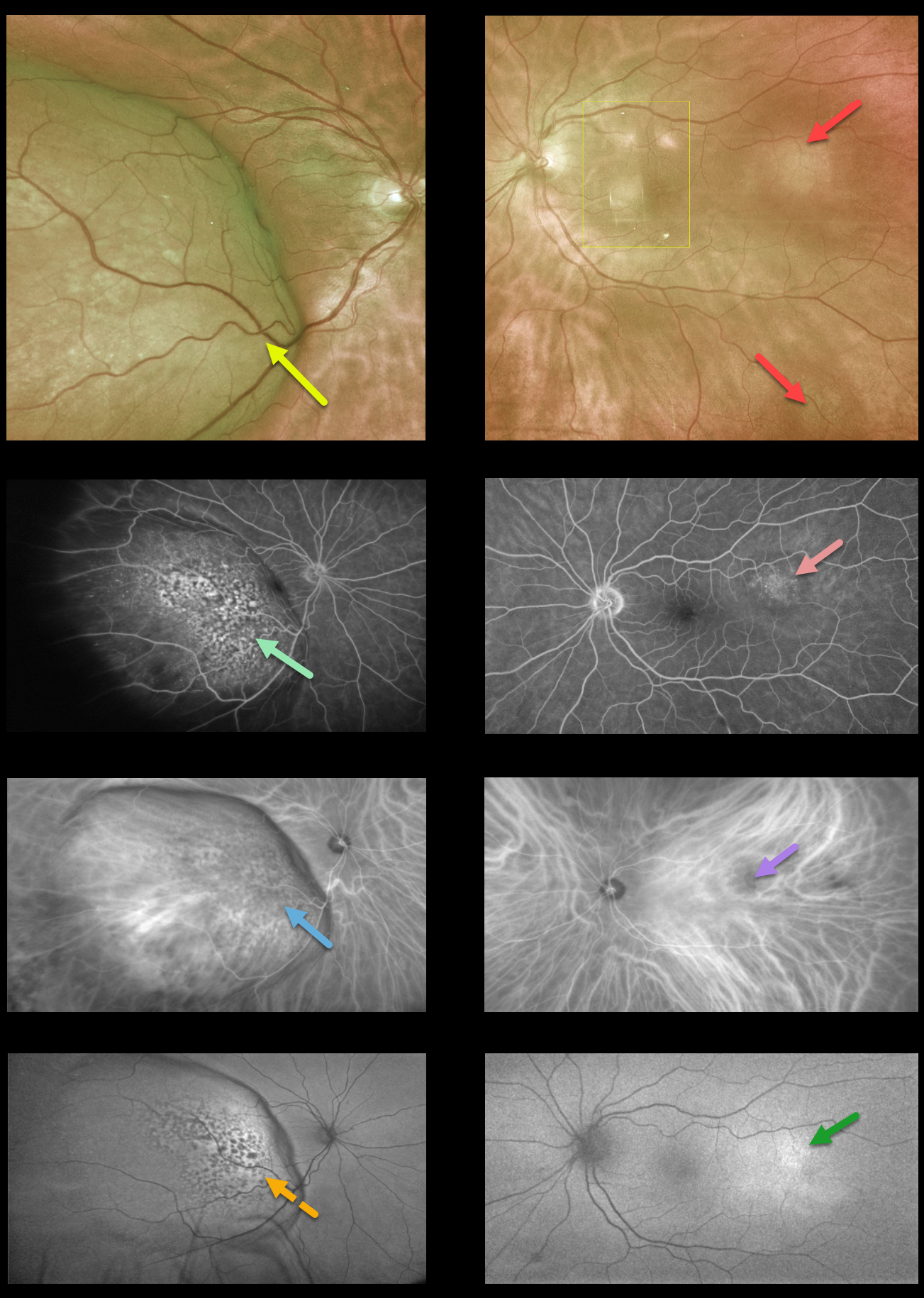
This 74YO female presented with a 2-week history of progressive vision loss in her right eye. Vision was 20/200 OD and 20/20 OS.
Optos imaging shows a large, creamy-colored temporal choroidal lesion extending into the right temporal macula. The fovea is markedly elevated and tilted along the nasal edge of the tumor. Two smaller and fainter amelanotic choroidal lesions are noted in the left eye (the lesions in the left macula are artifacts).
Fundus fluorescein angiography (FFA) shows patchy window defects over the nasal dome of the lesion in her right eye and faint staining from the temporal lesion in the left eye.
Indocyanine green angiography shows mostly hypofluorescence corresponding to the FFA window defects with some increased fluorescence from the intrinsic vasculature more temporally in the right eye, and hypofluorescence from the temporal lesion in the left eye.
Fundus autofluorescence (FAF) shows hypo-FAF corresponding to the FFA window defects in the right eye and hyperfluorescence from the temporal lesion in the left eye.
There was a known history of metastatic breast cancer, and a recent PET scan showed possible new lung metastases.
We are currently working with the patient’s oncologist for the next step forward, which will likely include external beam radiotherapy for a presumed choroidal metastasis in her right eye. Photodynamic therapy or transpupillary thermotherapy will be considered for the left eye.
Learning Points:
Although uveal melanoma is the most common primary intraocular malignancy, metastatic choroidal tumors are the most common intraocular malignancies. Lung and breast cancer are the most common sources.



