

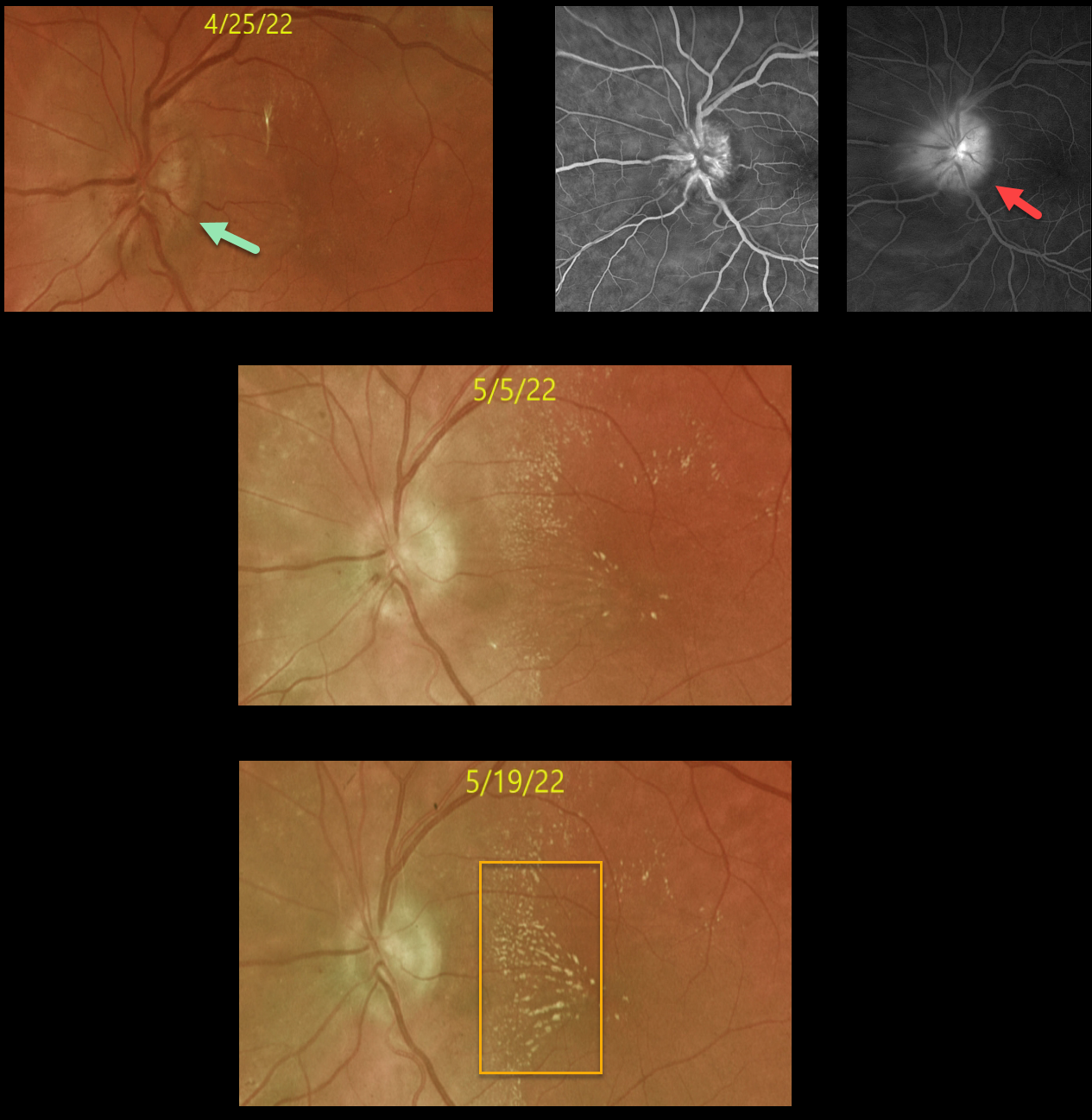
A 44YO female with type 2 diabetes presented on 4/25/22 with a 2-week history of severe vision loss in her left eye. Vision was counting fingers OS.
Ocular examinations were normal except for a markedly swollen left optic nerve. There were very faint, scattered, hard exudates.
OCT showed an average nerve fiber layer thickness of 370 microns (not shown). Fluorescein angiography shows diffuse optic nerve leakage.
Extensive bloodwork was negative, including Bartonella, syphilis, Lyme, Rocky Mountain spotted fever, CRP, ACE, and serum lysozyme. MRI scanning was also normal.
Over the next 3-4 weeks, the optic nerve edema resolved with the development of a nasal lipid star. Unfortunately, vision remained at counting fingers.
Learning Points:
The diagnosis for a unilateral swollen nerve is extensive. However, it becomes much smaller when associated with acute inner retinal ischemia (not seen in our case) and later lipid exudation within Henle’s layer (neuroretinitis), including cat scratch (Bartonella), Lyme disease, and syphilis.
When no underlying cause is found, the entity is called Leber’s idiopathic stellate neuroretinitis.



