

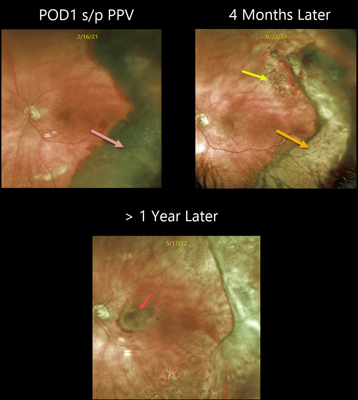
This 80YO female with a known history of dry AMD presented with a 3-week history of poor vision in her left eye. Vision was hand motion from a dense vitreous hemorrhage. B-scan ultrasonography showed a thickened temporal subretinal lesion without retinal detachment (not shown).
One day following vitrectomy, vision improved to 20/100. The temporal subretinal and RPE blood somewhat resembled a uveal malignant melanoma. However, the well-demarcated location and shape of the lesion, along with the posterior rim of subretinal blood, helped establish the diagnosis of a breakthrough vitreous hemorrhage from ectopic choroidal neovascularization (CNV), also known as peripheral exudative hemorrhagic chorioretinopathy (PEHCR).
Four months later, the resolved devitalized blood was turning yellow. The coarse subretinal pigmentation, most prominent superotemporally, is pathognomonic for prior subretinal blood.
She was subsequently lost to follow-up until she presented with a 6-month history of decreased vision. Although the PEHCR ridge continued to contract, vision was counting fingers due to a new macular neovascularization. Anti-VEGF therapy was started.
Learning Points:
PEHCR is an exudative process similar to wet AMD but located in the retinal periphery. Ectopic CNV can often masquerade as a choroidal mass or uveal melanoma.
It can also cause extensive choroidal/suprachoroidal, subretinal, and vitreous hemorrhage, more commonly in patients who are on blood thinners.
The subretinal blood often spontaneously regresses without treatment, although anti-VEGF injections can be used for symptomatic or increasing exudation.



