

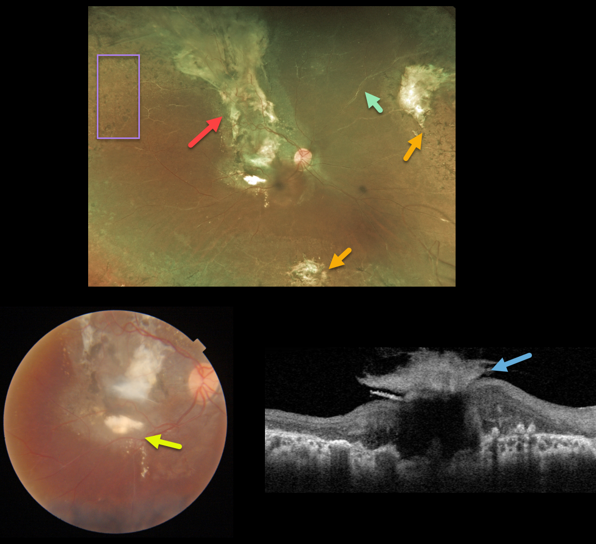
This 15YO female received prior thermal laser for Coats disease over 10 years earlier and was subsequently lost to follow-up. Right eye vision was counting fingers.
Optos color imaging shows subretinal scarring extending from the macula through the superior retinal periphery, nasal and inferior peripheral scarring, scattered peripheral retinal laser scarring, and sheathed vessels. There are no active Coats lesions.
Triton color imaging shows a magnified view of the macular scarring with some inferior macular subretinal lipid. Swept-source OCT shows a volcano-like vitreous component to the macular scar.
Learning Points:
Historically considered a unilateral disease, more recent ultra-widefield angiographic studies show subtle vascular changes, mostly in the temporal periphery (Brockmann et al., BJO 2021;105:1444-1453). Our patient’s left eye, however, was completely normal.



