

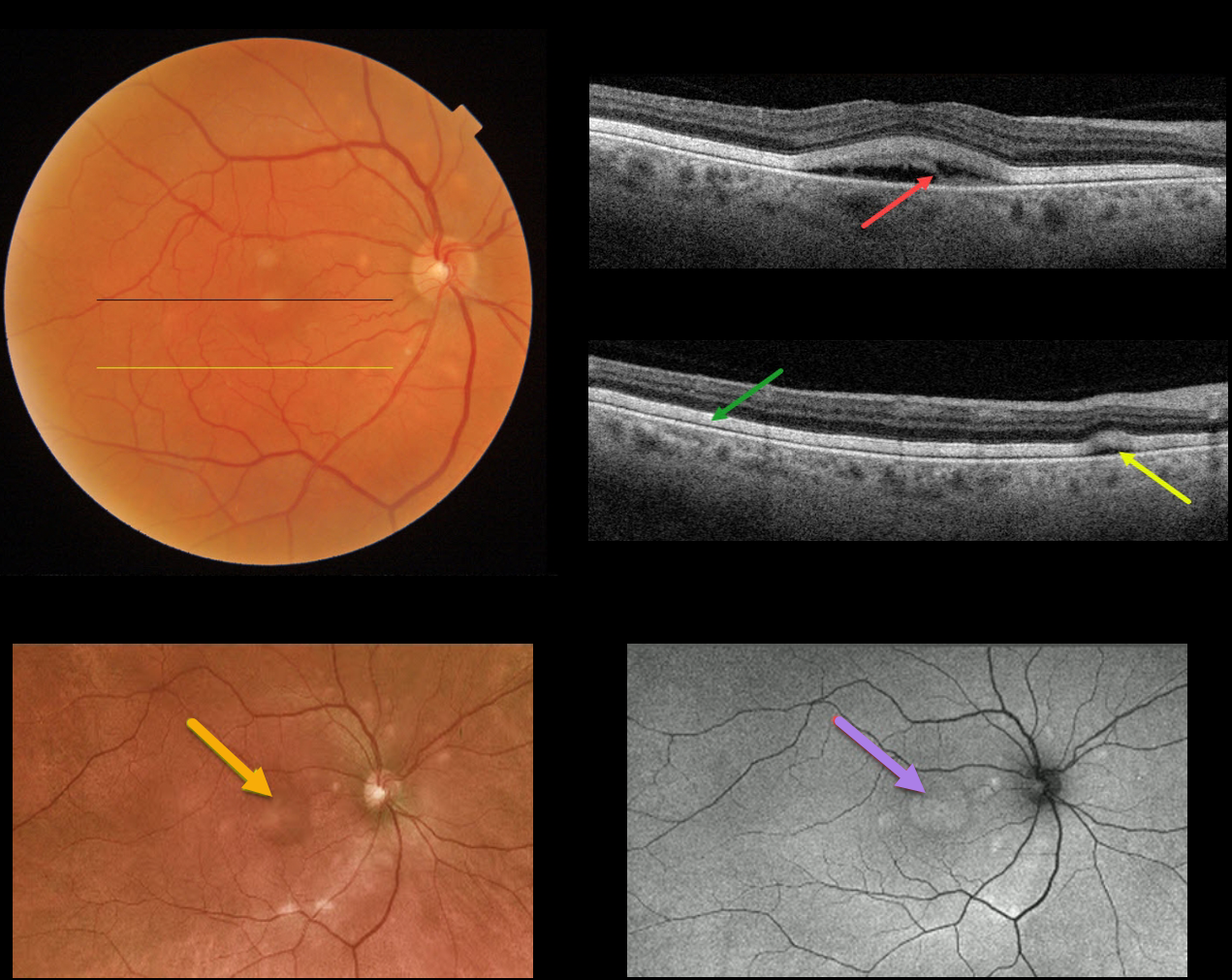
This 72YO male presented with recent difficulties with dark adaptation and 20/60 vision in his right eye. His left eye was enucleated 2 years earlier for a uveal malignant melanoma.
Triton color imaging shows multiple yellowish subretinal round lesions scattered around the optic nerve and macula.
Triton Swept Source OCT through the fovea shows a partially scrambled vitelliform lesion. An OCT B-scan through the inferior macula shows the outer retina diffusely infiltrated by hyperreflective vitelliform material, along with a small serous detachment.
Optos imaging shows an ultrawide view of the smaller and larger central vitelliform lesions, and fundus autofluorescence (FAF) shows hyper-FAF from the corresponding vitelliform lesions. ICG and FA were both normal.
Learning Points:
Acute exudative polymorphous vitelliform maculopathy (AEPVM) is a rare condition characterized by acute vision loss from multifocal serous retinal detachments that later progress to subretinal vitelliform lesions.
Initially, the serous detachments can mimic central serous retinopathy, including choroidal thickening. The condition is often self-limiting, and vision usually recovers within several months. The yellow vitelliform lesions may take 1-2 years to resolve completely.
These patients need a systemic workup for metastatic disease, since paraneoplastic retinopathy, including metastatic melanoma, can mimic idiopathic AEPVM.
Sadly, preliminary metastatic workup for this patient showed two possible liver metastases.



