

This 48YO male gave a history of lifelong poor vision in his left eye. Vision was 20/40 OD and 20/200 OS.
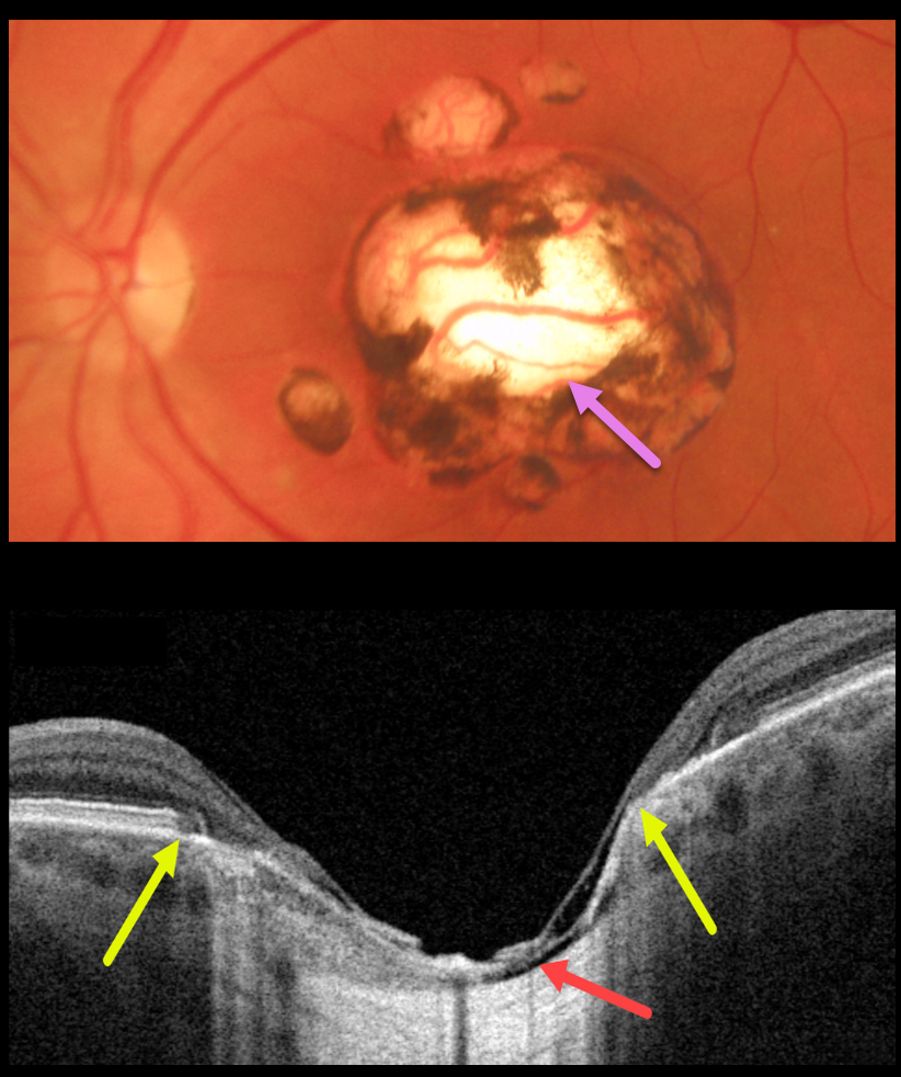
Triton color imaging shows a large, round, variably pigmented, excavated macular scar with a few smaller surrounding scars.
Swept-source OCT reveals a thinned atrophic retina and RPE overlying a shallow coloboma. Within the coloboma, the choroid is virtually absent, and the sclera is excavated. The yellow arrows mark the hyper-reflective plane of the surrounding intact retina and RPE. The right fundus was normal.
Learning Points:
For a great discussion on how to differentiate these infectious macular colobomatous lesions (congenital toxoplasmosis, Zika, and cytomegalovirus) from inherited dystrophies like North Carolina Macular Dystrophy (NCMD), see Kumar and Mahalingam AJO 2019;200:47-56.



