

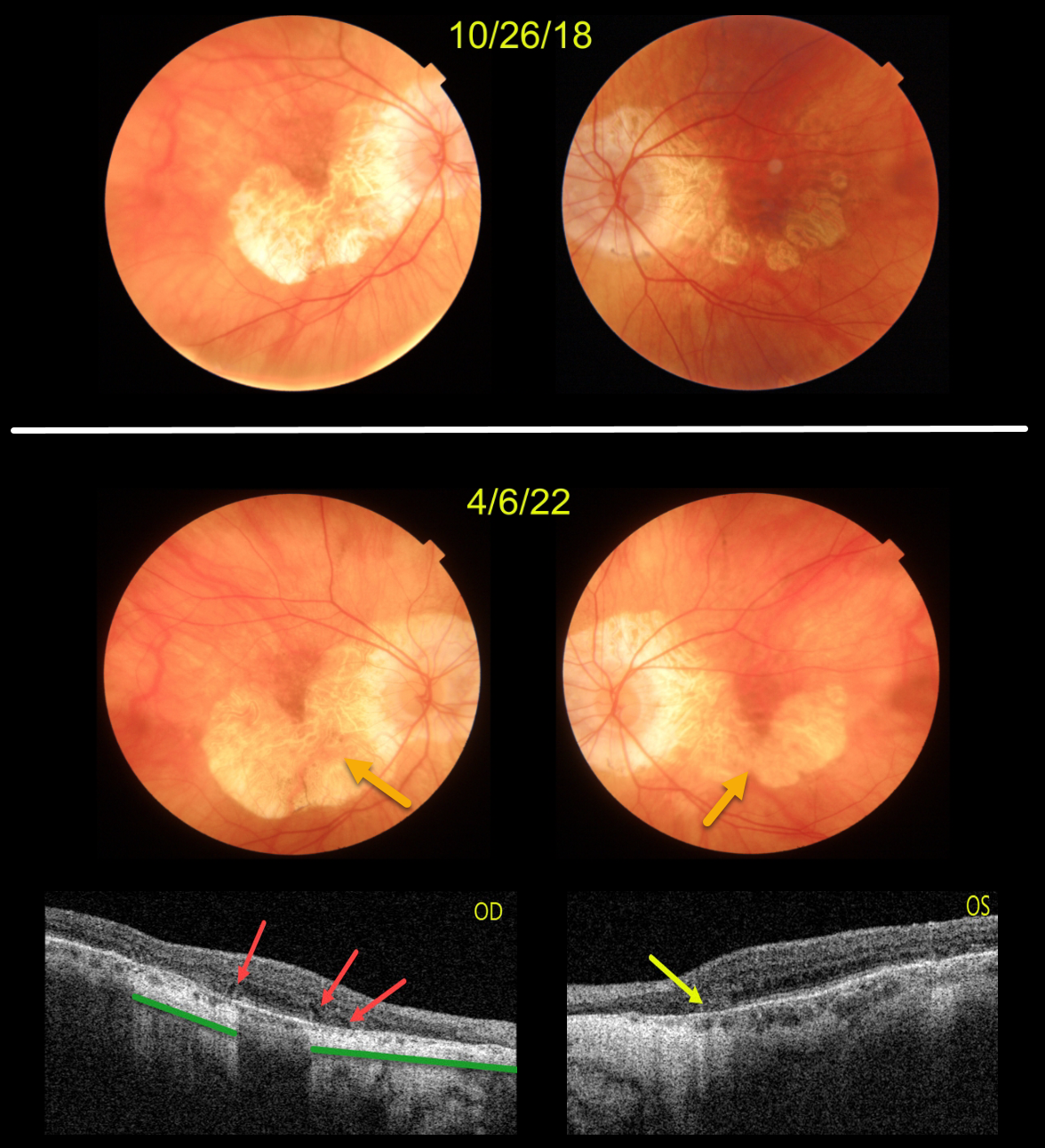
This patient originally presented in 2018 with vision of 20/40 OD and 20/50 OS. Areas of AMD-related macular atrophy (MA) skirted each macular center.
Four years later, vision had decreased to 20/100 OU due to progressive atrophy. Comparing the two images from 2018 to 2022, there is a bilateral linear enlargement for all margins of the atrophy, particularly in the left eye.
Triton swept-source OCT B-scans show classic findings for MA. The right eye has multiple wedge defects, hyporeflective wedges which develop at the boundaries of the degenerating outer plexiform layer in about 75% of MA eyes (Mones et al, Ophthalmology 2012;119:1412-1419). In addition, loss of the outer retina and RPE allows more OCT light to be transmitted into the choroid, resulting in distinct areas of choroidal and scleral hyperreflectivity.
There is a tiny area of outer retinal tubulation (ORT) nasally in the left eye, which represents photoreceptor and Muller cell degeneration (Dolz-Marco et al, Ophthalmology 2017;124:1353-1367). These ORTs have a hyper-reflective border with a central hyporeflective core.



