

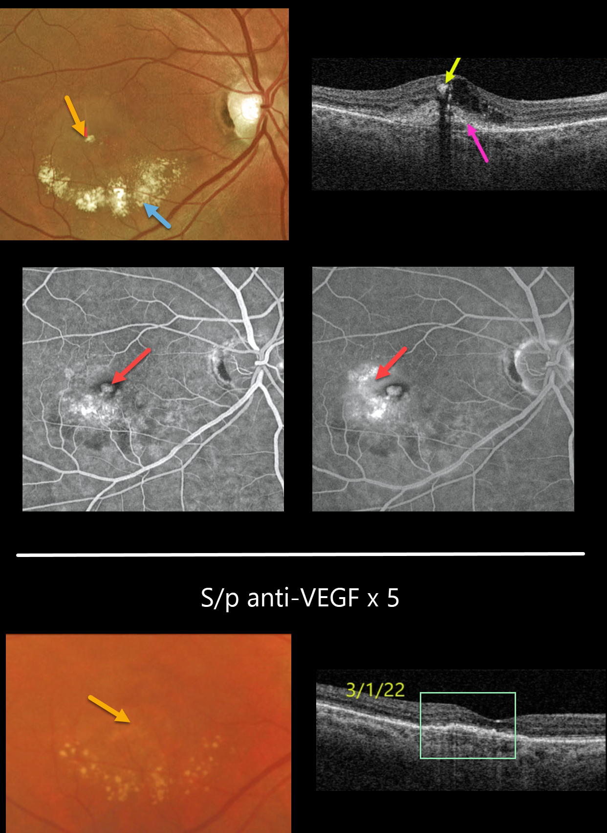
This 64YO female presented with this symptomatic unilateral exudative lesion in her right eye. Vision was 20/200.
Optos color imaging shows a small inner retinal yellow-white nodule with inferior subretinal lipid.
Triton swept-source OCT shows hyperreflectivity of this nodule with posterior shadowing, surrounding inner retinal fluid, and a hyperreflective macular neovascularization (MNV) located both below (type 1) and above (type 2) the RPE.
Optos fluorescein angiography shows a hyperfluorescent lesion with late leakage. The left eye showed mild macular drusen and pigmentary changes (not shown). This patient’s MNV is somewhat atypical given the extent of lipid exudation.
With monthly anti-VEGF therapy, the exudation resolved and vision improved to 20/60 following 5 intravitreal injections. The yellow nodular inner retinal lesion also completely resolved.
We therefore believe this most likely represented an unusual collection of inner macular lipids rather than a type 3 MNV.



