

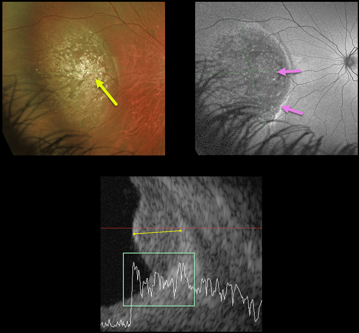
This 25YO male presented with this asymptomatic choroidal mass in his left eye. Vision was 20/20.
Optos color imaging shows a large, well-defined variably pigmented lesion with numerous overlying drusen in the nasal fundus. The lesion shows variable hypo- and hyper-autofluorescence.
B-scan ultrasonography shows moderate internal reflectivity. It measured 4 mm thick and 13.5 mm at its widest diameter.
We felt this lesion was a uveal malignant melanoma and referred him to an ocular oncologist for further evaluation and treatment.



