

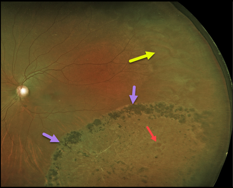
Years earlier, this 44YO female underwent laser demarcation of an asymptomatic inferotemporal rhegmatogenous retinal detachment (RRD). The prior detachment remains well demarcated, but the detachment itself has completely flattened.
Some of the causative lattice-related atrophic holes are seen within the area of prior detachment. White without pressure is also noted in the temporal periphery.
Learning Points:
Demarcation, rather than reattachment, of the retina is a valid option for select cases of RRD. When demarcating these detachments, it is critical that the retinopexy completely surrounds the subretinal fluid and that treatment extends to the ora; otherwise, the detachment can spread through the untreated retina.
Patients need to be followed postoperatively for life since the detachment can occasionally progress through the prior retinopexy.
Interestingly, we have seen that some of these detachments, as occurred in this case, spontaneously flatten following demarcation.
See Vrabec and Baumal, Ophthalmology 2000;107:1063-1067 for an excellent review regarding demarcation laser photocoagulation for selected macula-sparing retinal detachments.



