

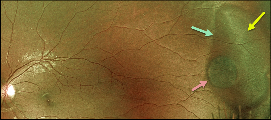
This 22YO female presented with a large temporal patch of dark without pressure (DWP), white without pressure (WWP), and a small round patch of congenital hypertrophy of the RPE (CHRPE). None of these findings is of concern, but we thought it was cool that all of them are associated with each other at this single location.
Learning Points:
WWP and DWP are relatively common benign peripheral findings most commonly found in darkly pigmented individuals. Typically, there are bilateral peripheral geographic areas of whitish (WWP) or dark (DWP) retinal discoloration.
WWP gets its name because the area is white without indentation, such as during scleral depression (white with pressure). It can sometimes be confused with retinal breaks or detachment.
The exact cause of this phenomenon is unknown. Historically, it was thought to be due to a benign vitreoretinal interface change, but more recent OCT findings show increased (WWP) or decreased (DWP) reflectivity in the outer retina (see Diaz et al., Retina 2014;34:1020-1021).
Although CHRPE lesions can enlarge with time (see Shields et al, Ophthalmology 2003;110:1968-1973), they carry virtually no malignant potential. Histologic RPE hypertrophy presents in several ways.
The typical isolated CHPRE lesions (this lesion) and bear tracks are not associated with intestinal polyps or cancer, and thus require no systemic evaluation. Only the multiple, small comet-shaped lesions are part of the familial polyposis spectrum.



