

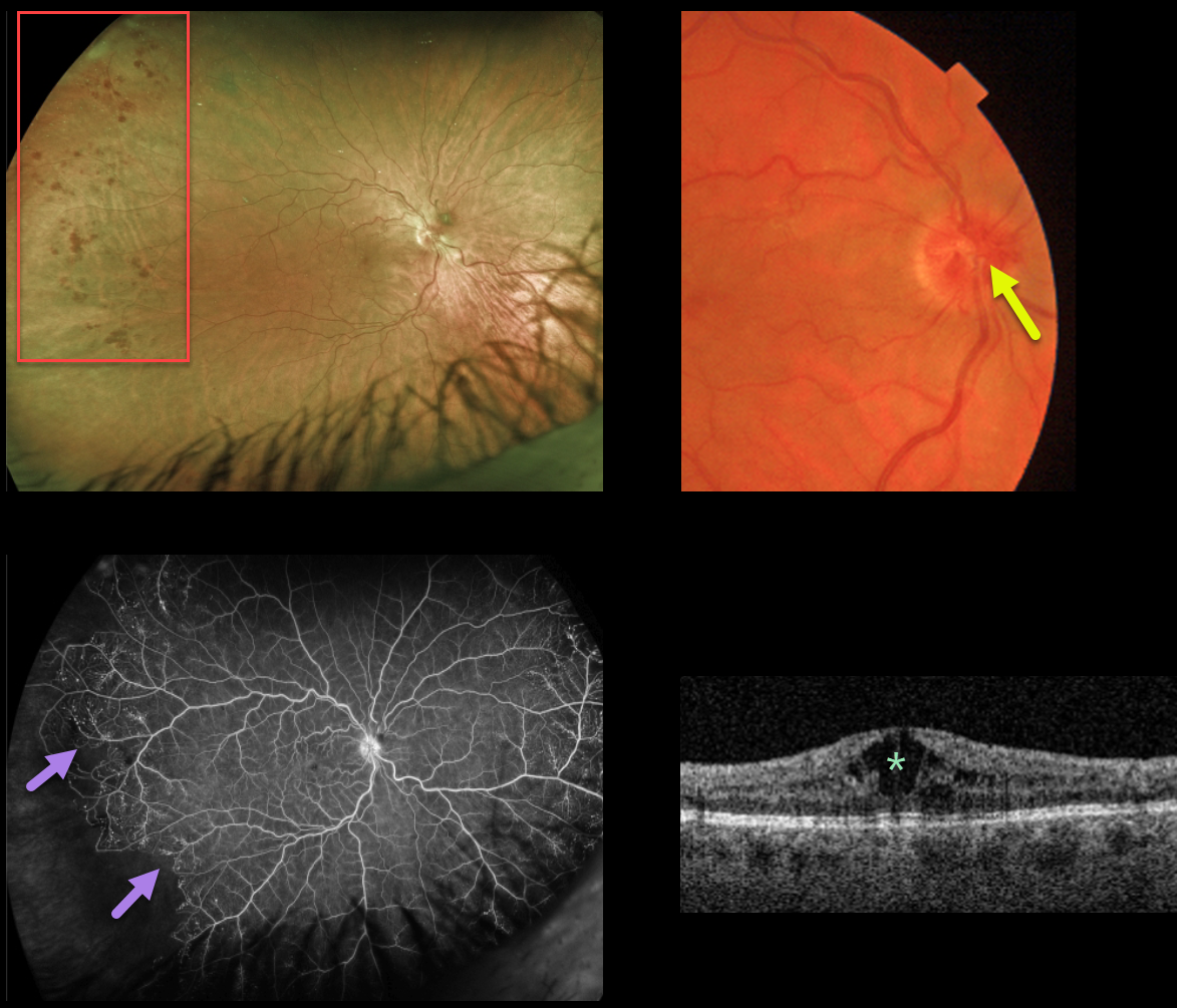
This 40YO female presented with 20/70 vision OD from a chronic central retinal vein occlusion (CRVO). The hemorrhages are virtually limited to the temporal periphery. Triton color imaging shows optic nerve collaterals.
Ultrawidefield Optos fluorescein angiography shows good posterior pole perfusion, nasal peripheral telangiectasia, and a sharp margin of infero-temporal peripheral ischemia.
Central cystoid edema is seen on swept-source OCT. Anti-VEGF injections were started for the macular edema.
Learning Points:
This sharply demarcated peripheral border of perfused ischemic retina is classically described in Eales and sickle retinopathy.
However, we have also found this to be a not uncommon finding in resolved CRVO, which has more classically been described as having vascular abnormalities extending outwards from the optic nerve. In this case, the optic disc collaterals point to the cause of the peripheral findings.
The Central Vein Occlusion Study (CVOS) defined ischemic CRVO as having at least 10 disc areas of angiographic retinal nonperfusion as determined by standard photographic views with the Canon (60 degrees) or Topcon (45 degrees) wide-angle fundus camera (Ophthalmology 1995;102:1434-1444).
Although clearly ischemic, this patient would have been classified by the CVOS as a perfused occlusion.
As with diabetic retinopathy (see Brucker, Retina 2021;41:461-463), ultrawidefield imaging and anti-VEGF injections have thrown a literal wrench into how we think about, classify, and treat retinal disorders.



