

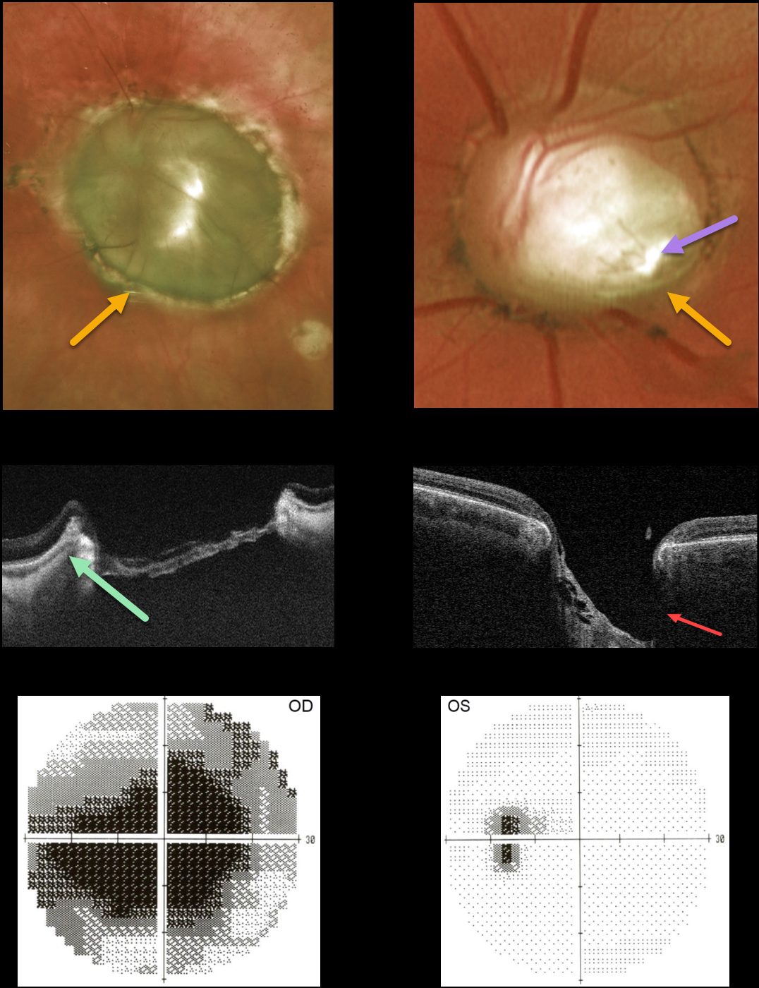
This 45YO female presented with bilateral optic nerve colobomas. Vision was hand motion OD and 20/25 OS.
Triton swept-source OCT shows a relatively featureless retina spanning the coloboma. The much smaller left nerve coloboma also contains an inferotemporal optic disc pit. The pit is also quite evident on OCT. The left nerve has a morning glory appearance with multiple vessels radiating outward from the peripheral disc margin, although there is no central glial tissue.
Visual fields show a large central defect OD with an enlarged blind spot OS.
Learning Points:
Optic nerve cavitary abnormalities can be unilateral or bilateral and result from incomplete closure of the embryonic fissure.
Our patient shows a spectrum of these abnormalities, including a huge optic disc coloboma OD, a much smaller disc coloboma OS with morning glory-like features, and an optic pit.
For a great review of cavitary optic nerve abnormalities, see Jain and Johnson, AJO 2014;158:423-435.



