

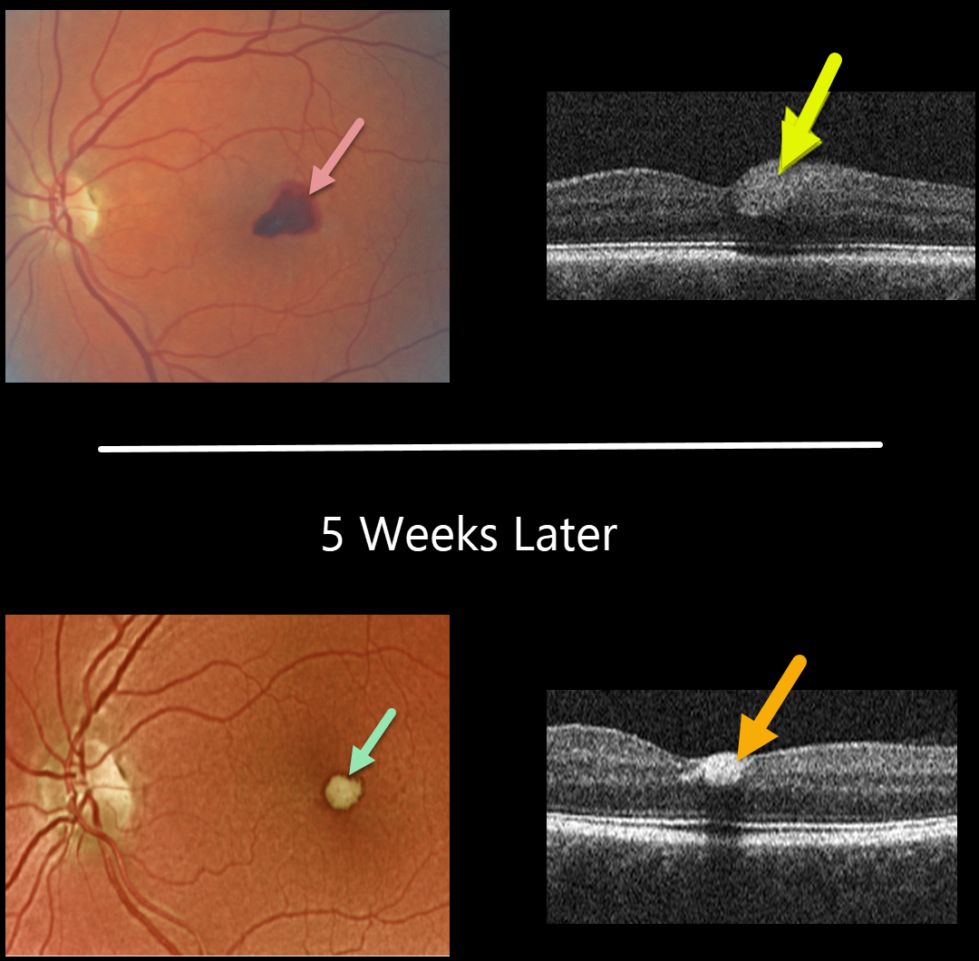
This 56YO female presented with acute vision loss in her left eye from Valsalva retinopathy. Vision was 20/100. A temporal foveal retinal hemorrhage is seen, located within the inner retina on OCT B-scan.
Five weeks later, the devitalized blood is contracting and turning yellow. The blood also became more hyperreflective on the OCT B-scan. All blood subsequently resolved with the return of normal vision.
Learning Points:
Valsalva hemorrhages are usually in the sub-internal limiting membrane (ILM) space, although this case was in the inner retina. These hemorrhages virtually always resolve without sequelae, although the sub-ILM blood can be drained into the inferior vitreous by creating a small opening in the ILM using either a thermal or Nd:YAG laser.



