

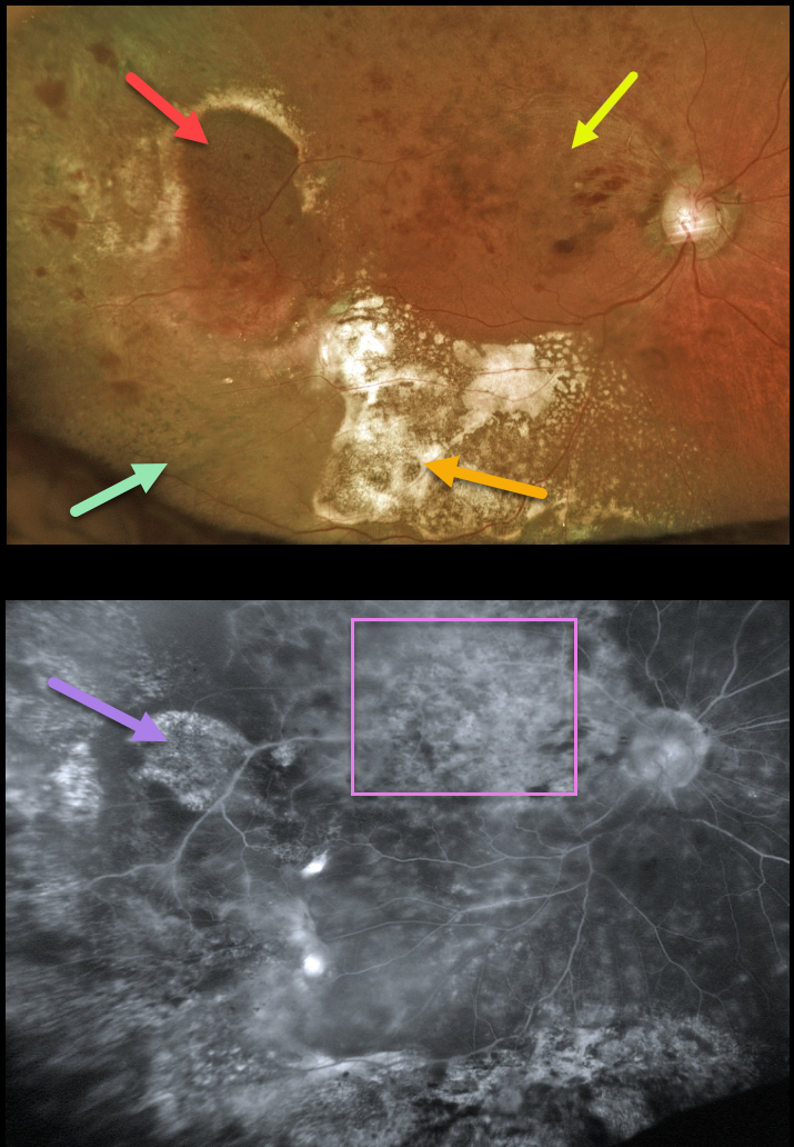
This 94YO female presented with ectopic choroidal neovascularization (CNV), also known as peripheral exudative hemorrhagic chorioretinopathy (PEHCR) in her right temporal periphery. Inferior to this lesion are clumps of subretinal pigment. Confluent subretinal lipid is also noted more inferiorly.
Vision was 20/400 due to macular edema from an unrelated major superotemporal branch retinal vein occlusion (BRVO). The inner retinal hemorrhages from the BRVO extend into the region of the ectopic subretinal lesion, somewhat confusing the clinical picture.
Fluorescein angiography shows variable diffuse staining of the ectopic CNV and secondary pigmentary changes, along with diffuse leakage from the BRVO.
Learning Points:
PEHCR is an exudative process similar to wet AMD but located in the retinal periphery. Ectopic CNV can often masquerade as a choroidal mass or uveal melanoma. It can also cause extensive choroidal/suprachoroidal, subretinal, and vitreous hemorrhage, more commonly in patients who are on blood thinners.
The subretinal blood often spontaneously regresses without treatment, although anti-VEGF injections can be used for symptomatic or increasing exudation. The inferior subretinal pigment in our patient is a classic finding for resolved subretinal blood.



