

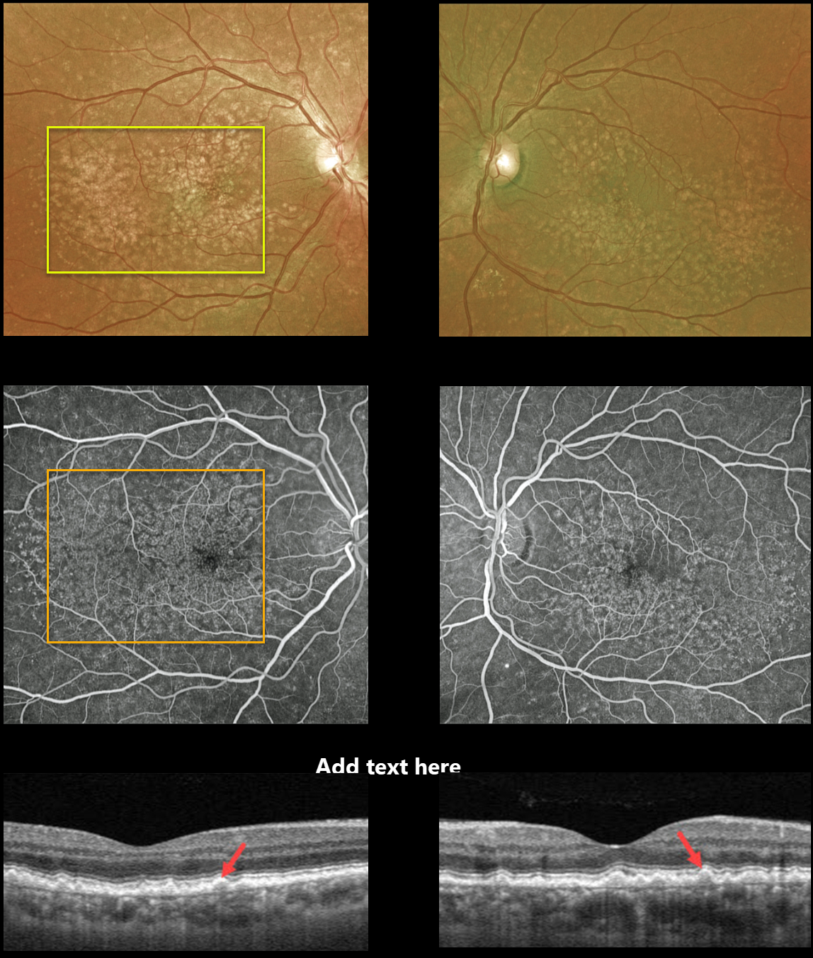
This 62YO male presented with 20/20 vision OU and asymptomatic macular drusen. Optos photos of both eyes show numerous scattered cuticular drusen throughout each macula.
Fluorescein angiography shows a dramatic bilateral starry sky pattern of innumerable hyperfluorescent drusen.
Triton swept-source OCT reveals multiple tooth-like cuticular drusen.
Learning Points:
Cuticular drusen, formerly known as basal laminar drusen, appear as numerous, round, yellow-white punctate accumulations located above Bruch’s membrane and below the RPE.
Originally described by Gass as a unique entity (AJO 1985;99;445-459), they are now thought to fall at least in part within the AMD spectrum. They are classically associated with avascular vitelliform lesions, but can also cause macular neovascularization.



