

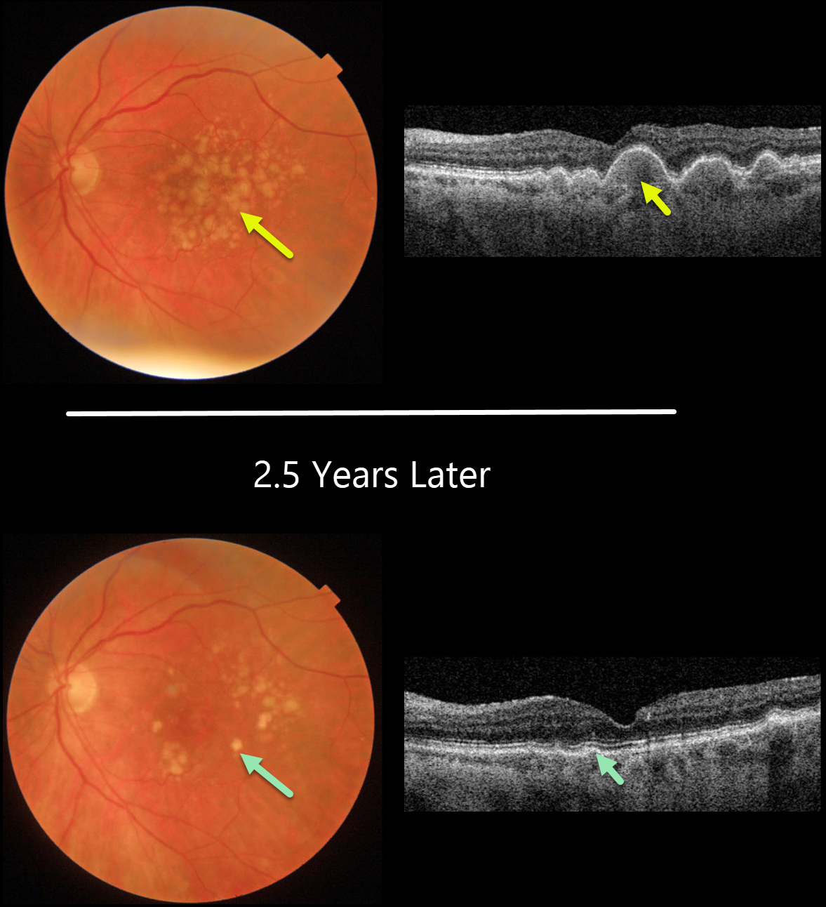
This 77YO female presented with diffuse, large, confluent soft drusen in both eyes (only left eye pictured). Vision was 20/50.
Two and a half years later, her macular examinations appeared quite different with marked drusen resolution. OCT shows near-complete flattening of all drusen, with irregularity of the outer retinal bands and RPE. Vision actually improved to 20/25.
Learning Points:
Drusen commonly remodel over time, and their collapse can lead to drusenoid atrophy of the RPE.
Although our patient’s outer retina is certainly abnormal on OCT, her clinical macular findings are remarkably benign given the extent of drusen several years ago.



