

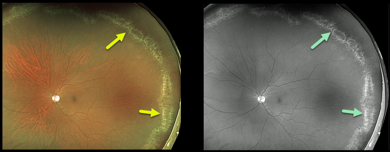
This 36YO male presented with extensive asymptomatic lattice degeneration seen on Optos color photography and Optos green channel.
The circumferential glistening lattice lesions are also called snail tracks. Treatment was not recommended.
Learning Points:
White, fibrosed vessels often crisscross the lesions, which gives rise to the term “lattice.” There is vitreous liquefaction overlying the thinned retina with a firm vitreo-retinal adhesion along the borders of the lesions. This firm vitreoretinal adhesion, along with the often-present atrophic holes, can predispose these eyes to retinal detachment.
Retinal detachment occurs in 1 in 10,000 people per year. Lattice is found in 10% of the general population, especially in high myopes. Since about one-third of patients with retinal detachment have lattice as the cause, the question always arises, especially in patients like this: Should prophylactic retinopexy be performed? The short answer is that asymptomatic lattice generally should not be treated.



