

This 35YO female presented with a 2-week history of severe bilateral vision loss, photophobia, and red eyes. There was no significant past medical history.
Vision was counting fingers OU. There was moderate bilateral conjunctival injection, severe cells with moderate flare, moderate fibrin, and 360-degree posterior synechiae. Topical cycloplegia and steroids were prescribed.
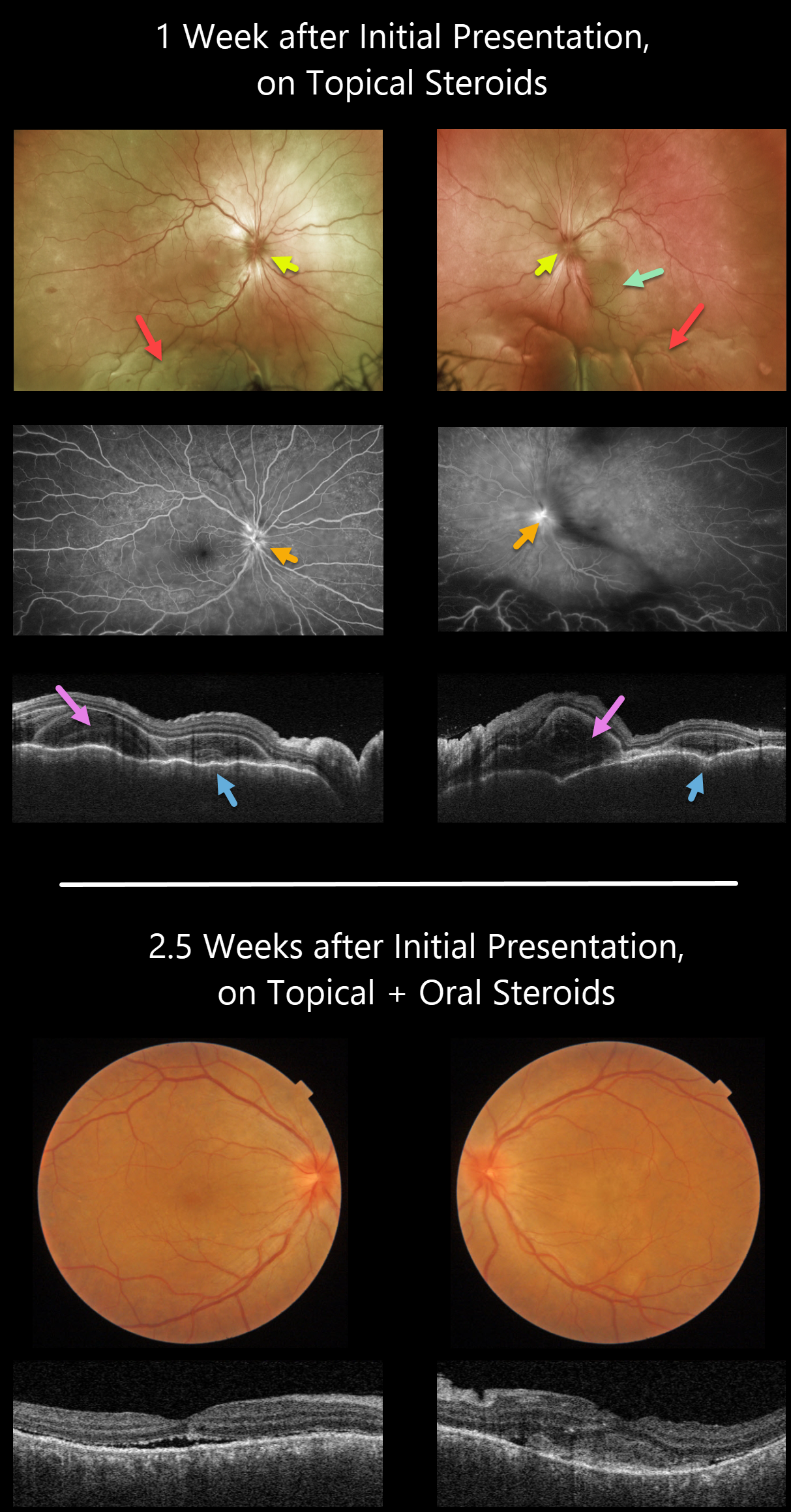
One week later, she was referred to our retinal service. There was marked improvement of her anterior uveitis and the posterior synechiae were mostly broken throughout. There were moderate bilateral vitreous cells, markedly swollen optic nerves, irregular diffuse macular exudative fluid with inferior exudative retinal detachments.
Fluorescein angiography showed bilateral optic nerve leakage. Triton swept-source OCT showed bilateral markedly thickened and rippled choroid with multiple bacillary pockets of outer retinal thickening and fluid. She started oral prednisone 80mg daily.
Around 10 days later, vision improved to 20/200 OU, with improvement in optic nerve edema, macular fluid, bacillary detachments, and choroidal thickening. The inferior exudative detachments were also resolving (not imaged).
Learning Points:
This case has many classic findings for Harada disease, including bilateral panuveitis, optic nerve swelling, thickened choroid, chorioretinal folds, and multifocal serous exudative retinal detachments.
Given the severity of the panuveitis and exudative subretinal fluid, we were surprised that angiography did not show the more classic multifocal subretinal leaks with pooling in the overlying serous detachments and bacillary cavities.
Patients with just ocular findings have Harada disease, whereas those with additional systemic findings (including vitiligo, poliosis, headache, vertigo, and hearing loss) have Vogt-Koyanagi-Harada disease.



