

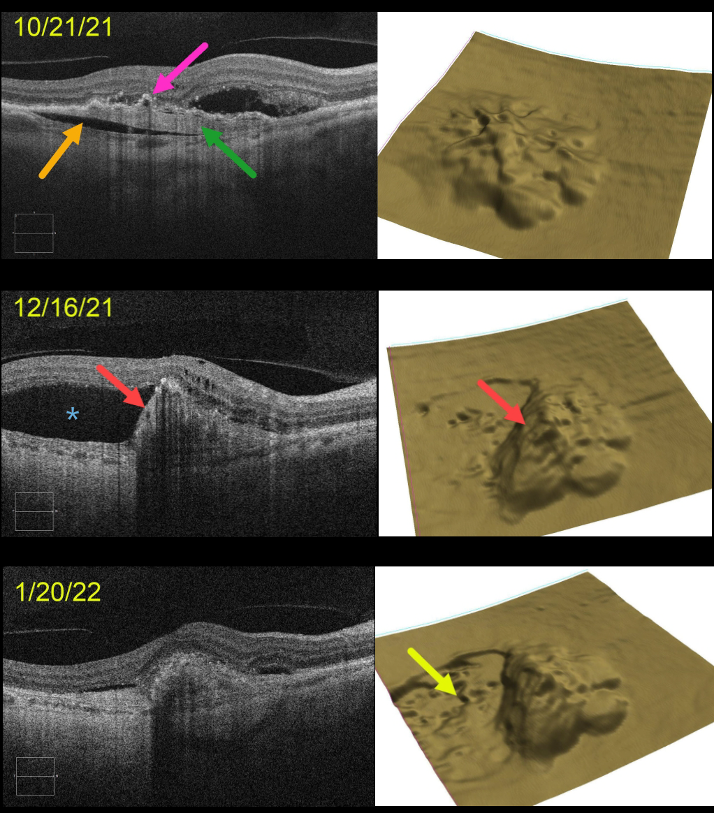
This 71YO female presented with a treatment-naive, age-related type 1 macular neovascularization (MNV). The already contracting MNV is elevated above the underlying choroid (prechoroidal cleft), and the overlying RPE is irregularly contracted and adherent to the MNV. The retinal pigment epithelium (RPE) map shows a 3-dimensional view of the RPE elevation. An anti-VEGF injection was given that day on 10/21/21.
On 12/16/21, vision was unchanged at 20/400 despite a new asymptomatic temporal RPE tear with marked overlying new subretinal fluid. An additional anti-VEGF injection was given.
On 1/20/22, vision was counting fingers. OCT scanning shows near-total resolution of all intra- and subretinal fluid. The RPE map nicely shows the crater of devoid RPE.
Learning Points:
A tear of the RPE is a fairly common occurrence in RPE detachment (PED) associated with type 1 MNV (located below the RPE). The neovascularization can spontaneously contract or involute following treatment. It then puts traction on the overlying RPE, which can then rip at the opposite edge of the PED.
This traction was already evident at our patient’s initial pre-injection visit, including the prechoroidal cleft, which is associated with a worse visual prognosis (see Kim et al, Retina 2017;37:2047-2055).
Like a cheap window blind, the torn RPE retracts and scrolls towards the MNV. This leaves a depigmented region where the RPE was and is now absent, along with a relatively straight, pigmented subretinal lesion extending from the scrolled RPE edge.



