

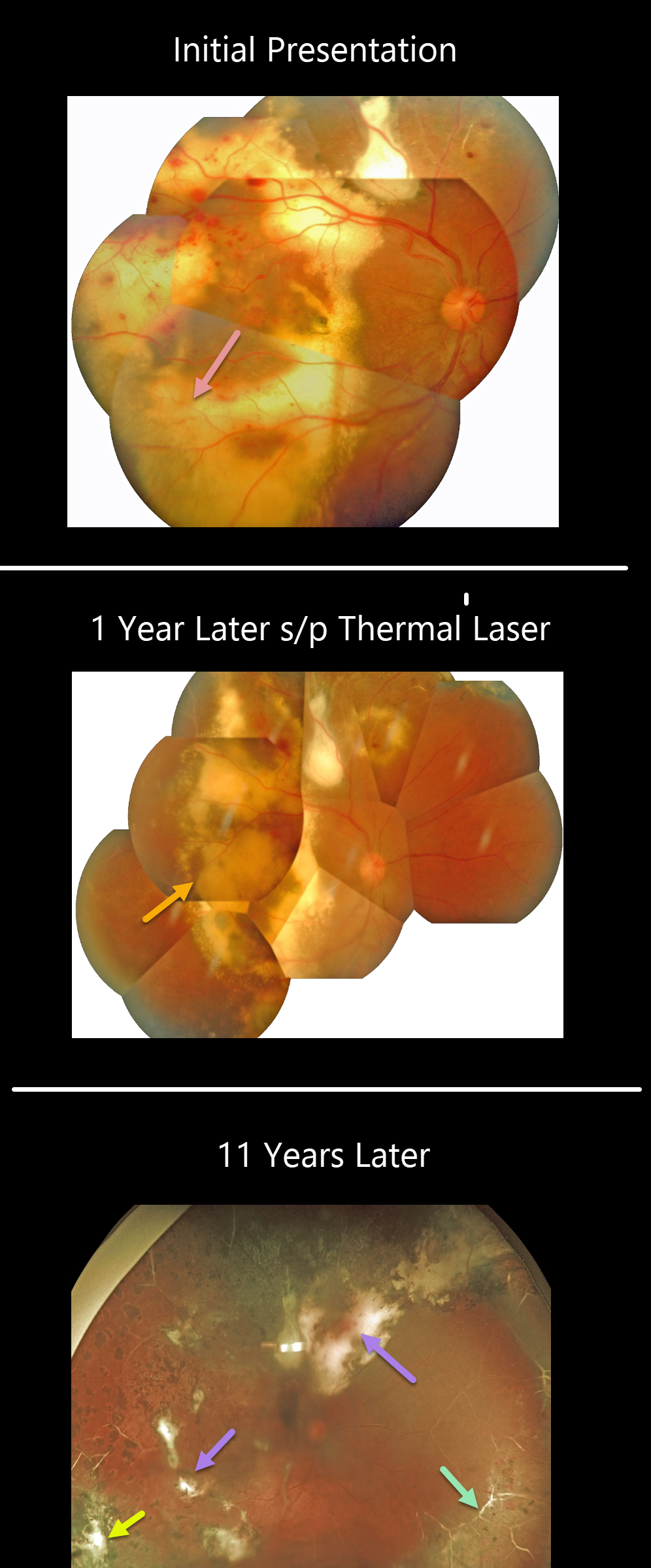
This patient was first seen in 2009 when he was 16 years old. Confluent subretinal lipid can be seen extending to the periphery. Several sessions of thermal laser were applied, and 1 year later, photos showed inactive Coats lesions with decreased subretinal lipid.
He was then lost to follow-up for 11 years when his vision was 20/400. Optos shows total resolution of all exudation and subretinal lipid with variable residual subretinal scarring. Peripheral scatter laser scarring is noted. The peripheral retinal vessels are white. There are no active Coats lesions.
Learning Points:
Historically considered a unilateral disease, more recent ultra-widefield angiographic studies show subtle vascular changes, mostly in the temporal periphery (Brockmann et al., BJO 2021;105:1444-1453).
Our patient’s left eye, however, was completely normal clinically and angiographically.



