

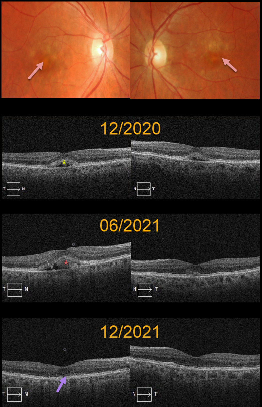
This 73YO female presented in 12/20 with bilateral central vitelliform lesions and more peripheral yellowish macular lesions, consistent with a probable macular dystrophy.
OCT shows bilateral subretinal hyperreflective lesions with underlying hyporeflectivity. She was asymptomatic with 20/25 vision OU. There was no family history of macular dystrophy. Genetic testing was negative, including for BEST1.
Six months later, the vitelliform lesion increased OD and decreased OS. Another six months later, the vitelliform lesions collapsed OU, and vision remained 20/25.
Learning Points:
Vitelliform lesions are commonly found in association with other disorders, including retinal dystrophies such as Best disease, macular dystrophies, cuticular drusen, AMD, and vitreomacular traction.
The yellow subretinal material is felt to be debris from shed photoreceptor outer segments. These lesions markedly autofluoresce on fundus autofluorescence imaging, which unfortunately, is unavailable for this patient.
A great reference for learning more about AVLs is Freund et al, Retina 2011;31:13-25.



