

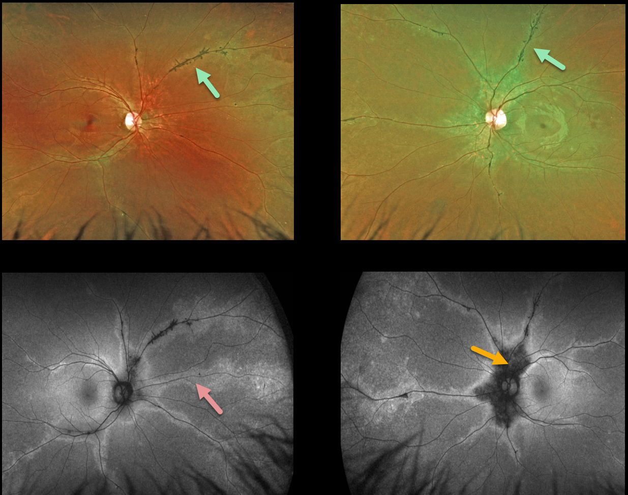
This 24YO female presented with 20/20 vision bilaterally and no visual symptoms. Optos ultrawidefield imaging shows normal central maculas with intraretinal pigment migration and depigmented RPE along the retinal veins. These changes extend from the peripapillary retina into the retinal peripheries.
Fundus autofluorescence (FAF) best highlights these changes with peripapillary hypo-FAF and paravenous hyper-FAF.
Our patient underwent Spark genetic testing. Results showed a pathogenic TNEM216 variant, which is associated with autosomal recessive Joubert Syndrome. However, she had absolutely no signs or symptoms of this syndrome.
Learning Points:
Pigmented paravenous chorioretinal atrophy (PPCRA) is a rare condition characterized by paravenous pigment clumps with peripapillary and radial zones of RPE atrophy.
It is bilateral, often asymmetric, with various causes, including genetic and pseudo-PPCRA (inflammatory, infectious, or unknown).
Patients tend to have some symptoms, usually nyctalopia, but most retain good vision, especially those without macular involvement.
The underlying pathophysiology is likely a primary choroidopathy that causes overlying RPE and retinal changes. The chorioretinal changes are best visualized with FAF. For a great clinical review, see Lee et al, AJO 2021;224:120-132.



