

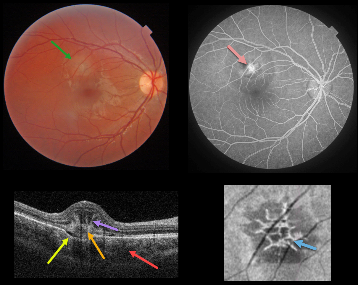
This healthy 14YO female presented with acute vision loss in her right eye. Vision was 20/40. Triton color photography shows a depigmented subretinal lesion in the superior macula.
Swept-source OCT shows a constellation of fascinating findings. A type 2 macular neovascularization is seen overlying a choroidal excavation. The choroid is thickened.
The most dramatic findings are the unusual linear, radiating, hyperreflective lesions extending anteriorly from the MNV, also known as the “pitchfork sign.” En face OCT through this lesion shows a spiky, wreath-like pattern of hyperreflectivity.
The MNV was confirmed on fluorescein angiography. Intravitreal anti-VEGF was begun.
Learning Points:
The pitchfork sign was originally described by Hoang et al (Retina 2013;33:1049-1055) as a unique OCT finding in inflammatory MNV. Falavarajani et al more recently noted that this finding is also associated with non-inflammatory MNV (Ophthalmic Surg Lasers Imaging Retina 2019;50:719-725). The pathogenesis of the avascular pitchfork spikes is unknown, but may be related to outer retinal traction or Müller cell activation.
Focal choroidal excavation, originally described by Jampol et al in 2006, is likely part of the pachychoroid spectrum and is associated with type 2 MNV. An excellent review is by Chung et al (Retina 2017;37;199-221).



