

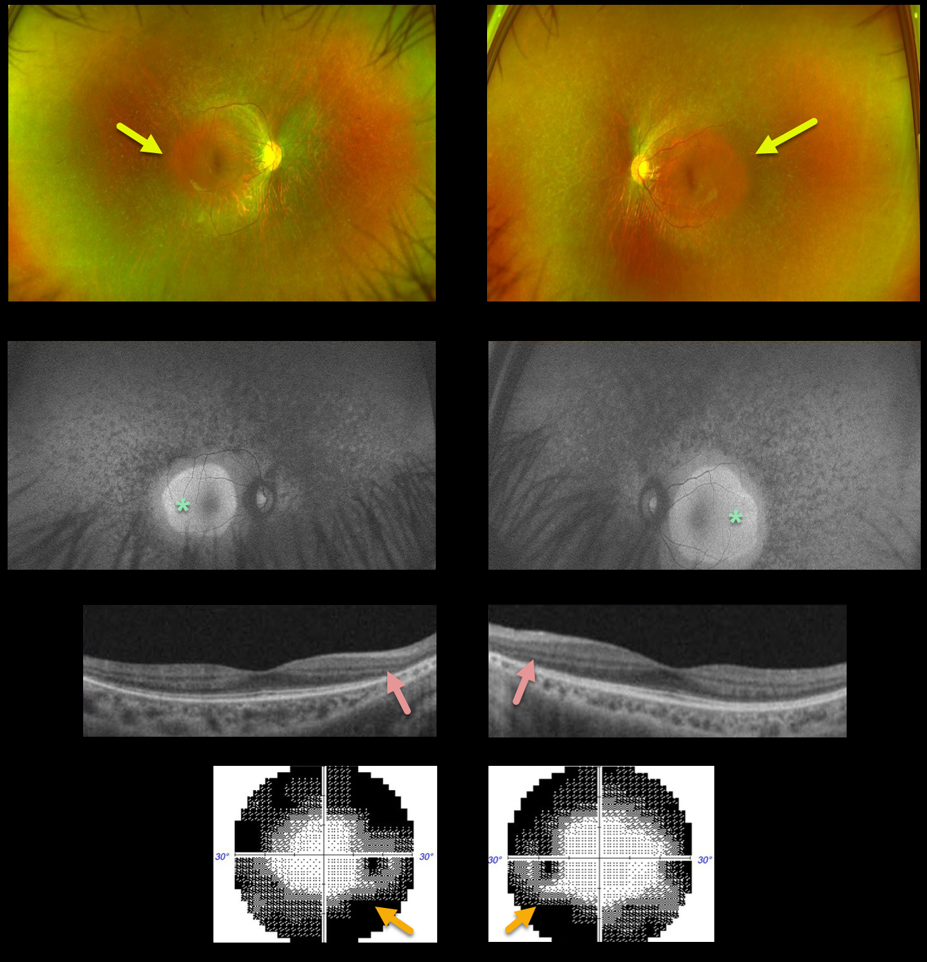
This 25YO male presented with complaints of nyctalopia and peripheral vision loss. Vision was 20/25 OU.
Optos color imaging shows a diffuse moth-eaten appearance to the RPE, sparing each macula. Minimal bone spicules are noted. Fundus autofluorescence (FAF) shows macular hyperautofluorescence.
OCT scanning shows peripheral outer retinal thinning. Visual field testing shows classing bilateral constriction. Genetic testing showed no pathogenic mutations.
Learning Points:
Patients with retinitis pigmentosa (RP) classically have pale optic nerves, narrowed retinal blood vessels, variable outer retinal and RPE loss, and intraretinal pigment migration (bone spicules) due to loss of the inhibitory effect of the degenerated outer segment,s allowing the RPE to migrate along the retinal capillaries. In real life, patients often lack all these features.
As in this case, intraretinal pigment migration may be minimal or absent but often appears over time (see Takahashi et al, AJO 2018;195:176-180).
A truly normal fundus in a patient with RP is extremely unusual, as there are usually very subtle clinical and FAF findings.



